. 2011;13(6):R117.
doi: 10.1186/bcr3060.

A different immunologic profile characterizes patients with HER-2-overexpressing and HER-2-negative locally advanced breast cancer: implications for immune-based therapies

Affiliations

A different immunologic profile characterizes patients with HER-2-overexpressing and HER-2-negative locally advanced breast cancer: implications for immune-based therapies

Elena Muraro et al. Breast Cancer Res. 2011.

Abstract

Introduction: The clinical efficacy of trastuzumab and taxanes is at least partly related to their ability to mediate or promote antitumor immune responses. On these grounds, a careful analysis of basal immune profile may be capital to dissect the heterogeneity of clinical responses to these drugs in patients with locally advanced breast cancer undergoing neoadjuvant chemotherapy.

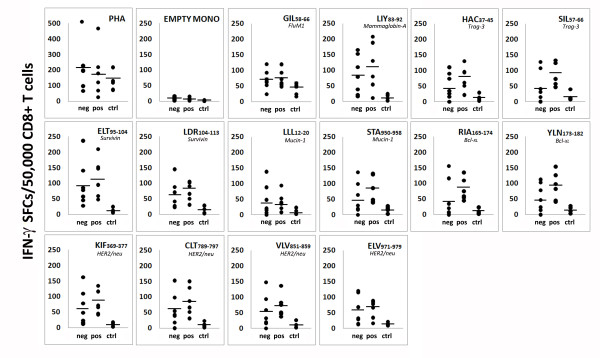
Methods: Blood samples were collected from 61 locally advanced breast cancers (36 HER2- and 25 HER2+) at diagnosis and from 23 healthy women. Immunophenotypic profiling of circulating and intratumor immune cells, including regulatory T (Treg) cells, was assessed by flow cytometry and immunohistochemistry, respectively. Serum levels of 10 different cytokines were assessed by multiplex immunoassays. CD8+ T cell responses to multiple tumor-associated antigens (TAA) were evaluated by IFN-γ-enzyme-linked immunosorbent spot (ELISPOT). The Student's t test for two tailed distributions and the Wilcoxon two-sample test were used for the statistical analysis of the data.

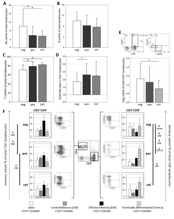
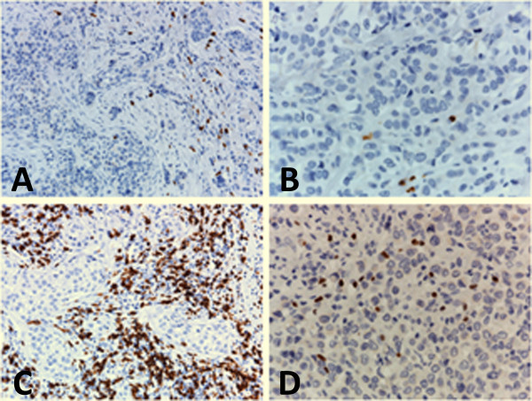
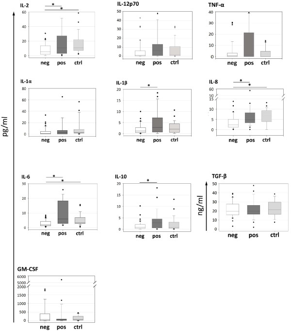
Results: The proportion of circulating immune effectors was similar in HER2+ patients and healthy donors, whereas higher percentages of natural killer and Treg cells and a lower CD4+/CD8+ T cell ratio (with a prevalence of naïve and central memory CD8+ T cells) were observed in HER2- cases. Higher numbers of circulating CD8+ T cells specific for several HLA-A*0201-restricted TAA-derived peptides were observed in HER2+ cases, together with a higher prevalence of intratumor CD8+ T cells. Serum cytokine profile of HER2+ patients was similar to that of controls, whereas HER2- cases showed significantly lower cytokine amounts compared to healthy women (IL-2, IL-8, IL-6) and HER2+ cases (IL-2, IL-1β, IL-8, IL-6, IL-10).

Conclusions: Compared to HER2- cases, patients with HER2-overexpressing locally advanced breast cancer show a more limited tumor-related immune suppression. This may account for the clinical benefit achieved in this subset of patients with the use of drugs acting through, but also promoting, immune-mediated effects.


Figures

Figure 1
Figure 1
Immunophenotypic characterization of peripheral blood lymphocytes. Percentages of (a) natural killer cells (NK; CD3-CD16+CD56+; neg = 20, pos = 17, ctrl = 17), (b) B cells (CD3-CD19+; neg = 14, pos = 15, ctrl = 15), (c) T lymphocytes (CD3+CD19-; neg = 20, pos = 17, ctrl = 17), (d) CD4+/CD8+ ratio (CD3+CD4+/CD3+CD8+; neg = 20, pos = 17, ctrl = 17), and (e) regulatory T cells (Treg; CD3+CD4+CD25highCD127lowFoxP3+; neg = 20, pos = 15, ctrl = 17) assessed in HER2- (neg), HER2-overexpressing (pos) patients and age-matched healthy women (ctrl). (f) Differentiation (memory) status of CD3+CD4+ and CD3+CD8+ lymphocytes was investigated through CCR7 and CD45RA expression (neg = 12; pos = 10; ctrl = 10). Statistical analysis was performed with t Student test. *, P<0.05 (refer to text for exact P value). HER-2, human epidermal growth factor receptor-2.
Figure 2
Figure 2
Composite figure showing CD8 and FoxP3 expression in lymphoid cells infiltrating representative HER2- or HER2+ breast carcinomas. (a) Few CD8+ cells are present within the tumor, and infiltrate tumor nests in a HER2- case. (b) Some FoxP3+ cells infiltrate a HER2- breast carcinoma. (c) CD8+ cells are numerous and surround tumoral cords in a HER2+ breast carcinoma. (d) Higher numbers of FoxP3+ cells infiltrate a HER2+ case. (a to d) Immunohistochemical stain; paraffin section; Hematoxylin counterstain; (a and c) 20× original magnification, (b and d) 40× magnification. HER2: human epidermal growth factor receptor-2.
Figure 3
Figure 3
CD8+ T cell responses to multiple breast cancer-associated antigenic epitopes assessed by IFN-γ-ELISPOT (interferon-γ-Enzyme Linked Immunosorbent Spot). All tests were performed using CD8+ purified T cells as effectors and autologous peptide-loaded monocytes as antigen presenting cells (APCs; effector:target ratio of 1:1). The number (enumerated as SFC, spot forming cells) of TAA-specific (or FluM1-specific, flu matrix protein1-derived epitope) circulating CD8+ T cells was investigated in HER2- (neg = 7) and HER2+ (pos = 6) breast cancer patients, whereas antigen-specific responses of healthy women were used as controls (ctrl = 5). PHA-loaded and empty monocytes (EMPTY MONO) were used as positive and negative controls, respectively. For peptides amino acid sequences, refer to Table 2. HER-2, human epidermal growth factor receptor-2; TAA, tumor-associated antigens.
Figure 4
Figure 4
Serum cytokine profile. Interleukin (IL)-2, IL-12p70, IL-1α, IL-1β, IL-8, IL-6, IL10 (neg = 36, pos = 25, ctrl = 23), Tumor necrosis factor-α (TNF-α; neg = 33, pos = 23, ctrl = 23), granulocyte-macrophage colony-stimulating factor (GM-CSF; neg = 33, pos = 23, ctrl = 19) and transforming growth factor-β (TGF-β; neg = 27, pos = 21, ctrl = 9) levels were evaluated in serum samples from HER2- (neg), HER2+ (pos) patients and age-matched healthy women (ctrl). Statistical analysis was performed with the Wilcoxon two-sample test. *, P<0.05 (refer to text for exact P value). HER2, human epidermal growth factor receptor-2.

References

    1. Mauri D, Pavlidis N, Ioannidis JP. Neoadjuvant versus adjuvant systemic treatment in breast cancer: a meta-analysis. J Natl Cancer Inst. 2005;97:188–194. doi: 10.1093/jnci/dji021. - DOI - PubMed
    1. Rouzier R, Mathieu MC, Sideris L, Youmsi E, Rajan R, Garbay JR, Andre F, Marsiglia H, Spielmann M, Delaloge S. Breast-conserving surgery after neoadjuvant anthracycline-based chemotherapy for large breast tumors. Cancer. 2004;101:918–925. doi: 10.1002/cncr.20491. - DOI - PubMed
    1. Ferriere JP, Assier I, Cure H, Charrier S, Kwiatkowski F, Achard JL, Dauplat J, Chollet P. Primary chemotherapy in breast cancer: correlation between tumor response and patient outcome. Am J Clin Oncol. 1998;21:117–120. doi: 10.1097/00000421-199804000-00003. - DOI - PubMed
    1. Borg A, Tandon AK, Sigurdsson H, Clark GM, Ferno M, Fuqua SA, Killander D, McGuire WL. HER-2/neu amplification predicts poor survival in node-positive breast cancer. Cancer Res. 1990;50:4332–4337. - PubMed
    1. Paik S, Hazan R, Fisher ER, Sass RE, Fisher B, Redmond C, Schlessinger J, Lippman ME, King CR. Pathologic findings from the National Surgical Adjuvant Breast and Bowel Project: prognostic significance of erbB-2 protein overexpression in primary breast cancer. J Clin Oncol. 1990;8:103–112. - PubMed

Publication types

MeSH terms