

Striatal amyloid plaque density predicts Braak neurofibrillary stage and clinicopathological Alzheimer's disease: implications for amyloid imaging
- PMID: 22112552
- PMCID: PMC3760731
- DOI: 10.3233/JAD-2011-111340
Striatal amyloid plaque density predicts Braak neurofibrillary stage and clinicopathological Alzheimer's disease: implications for amyloid imaging
Abstract
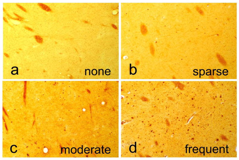
Amyloid imaging may revolutionize Alzheimer's disease (AD) research and clinical practice but is critically limited by an inadequate correlation between cerebral cortex amyloid plaques and dementia. Also, amyloid imaging does not indicate the extent of neurofibrillary tangle (NFT) spread throughout the brain. Currently, the presence of dementia as well as a minimal brain load of both plaques and NFTs is required for the diagnosis of AD. Autopsy studies suggest that striatal amyloid plaques may be mainly restricted to subjects in higher Braak NFT stages that meet clinicopathological diagnostic criteria for AD. Striatal plaques, which are readily identified by amyloid imaging, might therefore be used to predict the presence of a higher Braak NFT stage and clinicopathological AD in living subjects. This study determined the sensitivity and specificity of striatal plaques for predicting a higher Braak NFT stage and clinicopathological AD in a postmortem series of 211 elderly subjects. Subjects included 87 clinicopathologically classified as non-demented elderly controls and 124 with AD. A higher striatal plaque density score (moderate or frequent) had 95.8% sensitivity, 75.7% specificity for Braak NFT stage V or VI and 85.6% sensitivity, 86.2% specificity for the presence of dementia and clinicopathological AD (National Institute on Aging - Reagan Institute "intermediate" or "high"). Amyloid imaging of the striatum may be useful as a predictor, in living subjects, of Braak NFT stage and the presence or absence of dementia and clinicopathological AD. Validation of this hypothesis will require autopsy studies of subjects that had amyloid imaging during life.
Figures
References
-
- Villemagne VL, Pike KE, Darby D, Maruff P, Savage G, Ng S, Ackermann U, Cowie TF, Currie J, Chan SG, Jones G, Tochon-Danguy H, O’Keefe G, Masters CL, Rowe CC. Abeta deposits in older non-demented individuals with cognitive decline are indicative of preclinical Alzheimer’s disease. Neuropsychologia. 2008;46:1688–1697. - PubMed
-
- Reiman EM, Chen K, Liu X, Bandy D, Yu M, Lee W, Ayutyanont N, Keppler J, Reeder SA, Langbaum JB, Alexander GE, Klunk WE, Mathis CA, Price JC, Aizenstein HJ, DeKosky ST, Caselli RJ. Fibrillar amyloid-beta burden in cognitively normal people at 3 levels of genetic risk for Alzheimer’s disease. Proc Natl Acad Sci USA. 2009;106:6820–6825. - PMC - PubMed
-
- Pike KE, Savage G, Villemagne VL, Ng S, Moss SA, Maruff P, Mathis CA, Klunk WE, Masters CL, Rowe CC. Beta-amyloid imaging and memory in non-demented individuals: evidence for preclinical Alzheimer’s disease. Brain. 2007;130:2837–2844. - PubMed
-
- Mintun MA, Larossa GN, Sheline YI, Dence CS, Lee SY, Mach RH, Klunk WE, Mathis CA, DeKosky ST, Morris JC. [11C]PIB in a nondemented population: potential antecedent marker of Alzheimer disease. Neurology. 2006;67:446–452. - PubMed
Publication types
MeSH terms
Substances
Grants and funding
LinkOut - more resources
Full Text Sources
Medical
