. 2012 Mar;81(3):348-55.
doi: 10.1124/mol.111.076208. Epub 2011 Nov 23.

Desensitization and trafficking of μ-opioid receptors in locus ceruleus neurons: modulation by kinases

Affiliations

Desensitization and trafficking of μ-opioid receptors in locus ceruleus neurons: modulation by kinases

Seksiri Arttamangkul et al. Mol Pharmacol. 2012 Mar.

Abstract

The phosphorylation of μ-opioid receptors (MOPRs) by G protein-coupled receptor kinases (GRKs), followed by arrestin binding, is thought to be a key pathway leading to desensitization and internalization. The present study used the combination of intracellular and whole-cell recordings from rats and mice, as well as live cell imaging of Flag-tagged MOPRs from mouse locus ceruleus neurons, to examine the role of protein kinases in acute desensitization and receptor trafficking. Inhibition of GRKs by using heparin or GRK2-mutant mice did not block desensitization or alter the rate of recovery from desensitization. The nonselective kinase inhibitor staurosporine did not reduce the extent of [Met(5)]enkephalin (ME)-induced desensitization but increased the rate of recovery from desensitization. In the presence of staurosporine, ME-activated FlagMOPRs were internalized but did not traffic away from the plasma membrane. The increased rate of recovery from desensitization correlated with the enhancement in the recycling of receptors to the plasma membrane. ME-induced MOPR desensitization persisted and the trafficking of receptors was modified after inhibition of protein kinases. The results suggest that desensitization of MOPRs may be an early step after agonist binding that is modulated by but is not dependent on kinase activity.


Figures
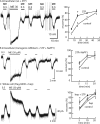
Fig. 1.
Fig. 1.
MOPR desensitization in the presence of protein kinase inhibitors. A, left, example trace showing desensitization and recovery from desensitization in a rat slice incubated with staurosporine (STP) (10 μM, 45 min); right, summarized results showing recovery from desensitization. B, left, example trace showing desensitization and recovery from desensitization in a GRK2as5-transgenic mouse slice incubated with staurosporine (10 μM, 45 min) and NaPP1 (1 μM); right, summarized results showing recovery from desensitization. C, left, example trace showing desensitization and recovery from desensitization in a FlagMOPR-transgenic mouse slice, recorded with heparin (hep) (1 mg/ml) in the pipette; right, summarized results showing recovery from desensitization for control, with heparin, and with heparin and staurosporine. *, P < 0.05, two-way ANOVA with Bonferroni post hoc test.
Fig. 2.
Fig. 2.
Staurosporine (STP)-induced qualitative changes in the distribution of internalized receptors from FlagMOPR-transgenic mice. Top, sequential images of an untreated slice; left, control staining with M1-A594 (45 min); middle, after treatment of the slice with ME (30 μM, 15 min); right, after treatment of the slice with calcium-free solutions. Bottom, sequential images of a staurosporine-treated slice; left, control staining with M1-A594 after incubation with staurosporine (10 μM, 45 min); middle, after treatment of the slice with ME (30 μM, 15 min); right, after treatment of the slice with calcium-free solutions.
Fig. 3.
Fig. 3.
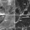
High-power images of staurosporine (STP)-treated cells from a FlagMOPR-transgenic mouse, illustrating receptor clustering in the cytoplasm near the plasma membrane. Top, images of a control slice; left, scanning infrared image showing the location of the plasma membrane (arrow); right, receptor staining with M1-A594 in the cytoplasm. Bottom, images of a staurosporine-treated slice; left, scanning infrared image showing the membrane of the cell; right, receptor staining with M1-A594. Arrows, locations of receptors close to the plasma membrane.
Fig. 4.
Fig. 4.
Staurosporine (STP)-induced MOPR clustering near the plasma membrane in a FlagMOPR-ArrKO mouse. Top, sequential images of an untreated slice; left, control staining with M1-A594 (45 min); middle, after treatment of the slice with ME (30 μM, 15 min); right, after treatment of the slice with calcium-free solutions. Bottom, sequential images of a staurosporine-treated slice; left, control staining with M1-A594 after incubation with staurosporine (10 μM, 45 min); middle, after treatment of the slice with ME (30 μM, 15 min); right, after treatment of the slice with calcium-free solutions.
Fig. 5.
Fig. 5.
Time course of FlagMOPR trafficking. Top, control; middle, after treatment with staurosporine (STP) (10 μM, 45 min); bottom, FlagMOPR-ArrKO mouse slice after incubation with staurosporine (10 μM, 45 min). Tg, transgenic.
Fig. 6.
Fig. 6.
Reinsertion of FlagMOPRs after internalization. A, top, left to right, control, after treatment with ME (30 μM, 15 min), after a washout period of 55 min, and after treatment with calcium-free solution; bottom, same experiment performed with a slice incubated with staurosporine (STP) (10 μM, 45 min). B, summarized results showing the amount of fluorescence remaining in slices after washout of ME (30 μM) for different periods. **, P < 0.01, two-way ANOVA with Bonferroni post hoc test.
Fig. 7.
Fig. 7.
Evidence that the p38MAPK inhibitor SB203580 (SB) does not block desensitization or change internalization but increases the extent of recovery from desensitization. A, example trace showing desensitization and recovery from desensitization in a slice incubated with SB203580 (1 μM, 45 min). *, P < 0.05, two-way ANOVA with Bonferroni post hoc test. B, summarized results showing the decline and recovery of hyperpolarization induced by ME (0.3 μM) after washout of ME (30 μM, 10 min). C, example images show sequential steps of MOPR internalization and reinsertion; left, control; middle, after treatment with ME (30 μM, 15 min) followed by washout; right, after stripping with calcium-free solutions.

References

    1. Armstrong N, Jasti J, Beich-Frandsen M, Gouaux E. (2006) Measurement of conformational changes accompanying desensitization in an ionotropic glutamate receptor. Cell 127:85–97 - PubMed
    1. Arttamangkul S, Quillinan N, Low MJ, von Zastrow M, Pintar J, Williams JT. (2008) Differential activation and trafficking of μ-opioid receptors in brain slices. Mol Pharmacol 74:972–979 - PMC - PubMed
    1. Arttamangkul S, Torrecilla M, Kobayashi K, Okano H, Williams JT. (2006) Separation of μ-opioid receptor desensitization and internalization: endogenous receptors in primary neuronal cultures. J Neurosci 26:4118–4125 - PMC - PubMed
    1. Bailey CP, Kelly E, Henderson G. (2004) Protein kinase C activation enhances morphine-induced rapid desensitization of μ-opioid receptors in mature rat locus ceruleus neurons. Mol Pharmacol 66:1592–1598 - PubMed
    1. Benovic JL, Stone WC, Caron MG, Lefkowitz RJ. (1989) Inhibition of the β-adrenergic receptor kinase by polyanions. J Biol Chem 264:6707–6710 - PubMed

Publication types

Substances