. 2012 Mar;132(3 Pt 1):642-9.
doi: 10.1038/jid.2011.377. Epub 2011 Nov 24.

IFN-γ, IL-21, and IL-10 co-expression in evolving autoimmune vitiligo lesions of Smyth line chickens

Affiliations

IFN-γ, IL-21, and IL-10 co-expression in evolving autoimmune vitiligo lesions of Smyth line chickens

Fengying Shi et al. J Invest Dermatol. 2012 Mar.

Abstract

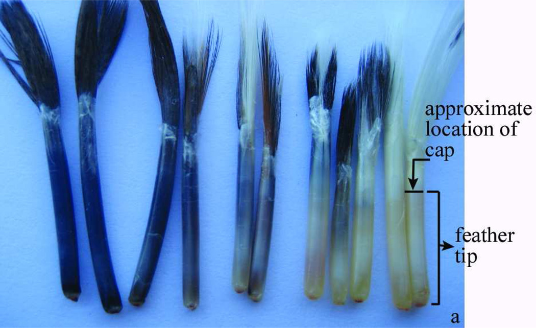
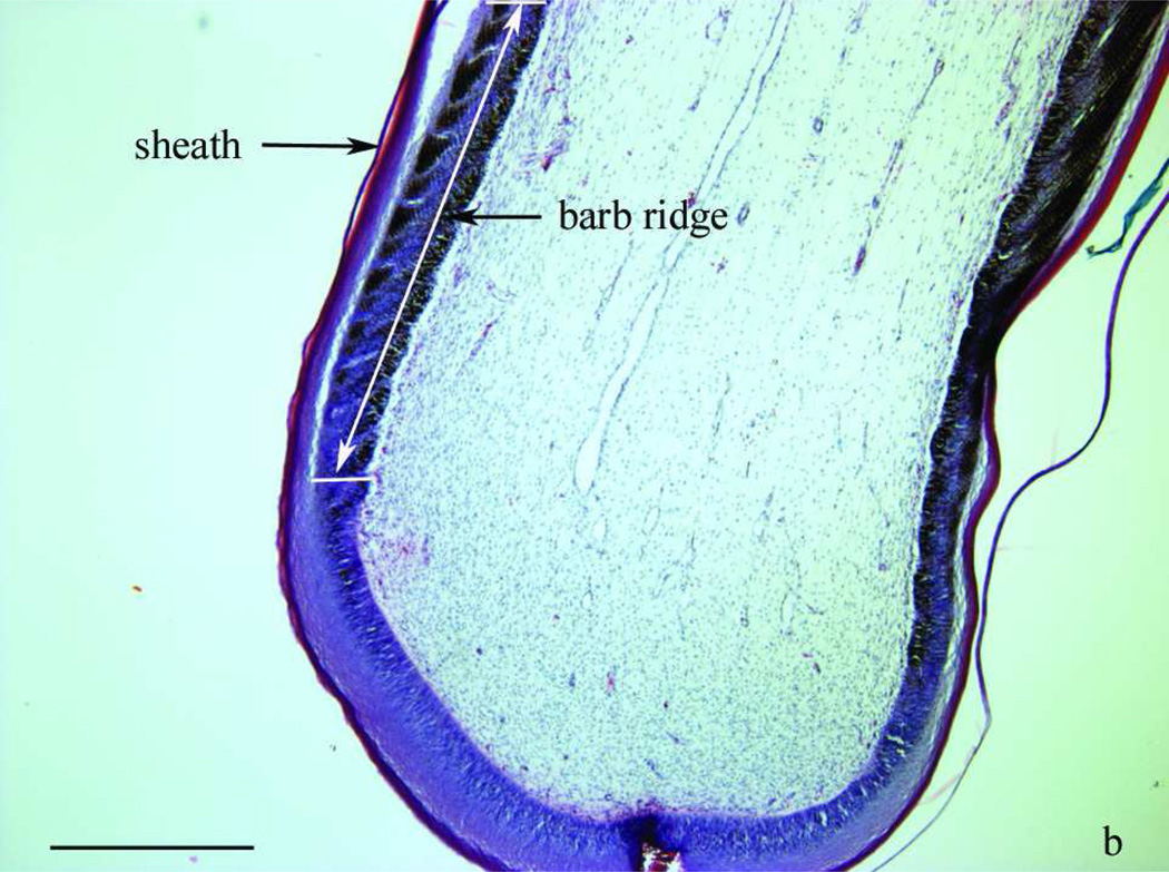
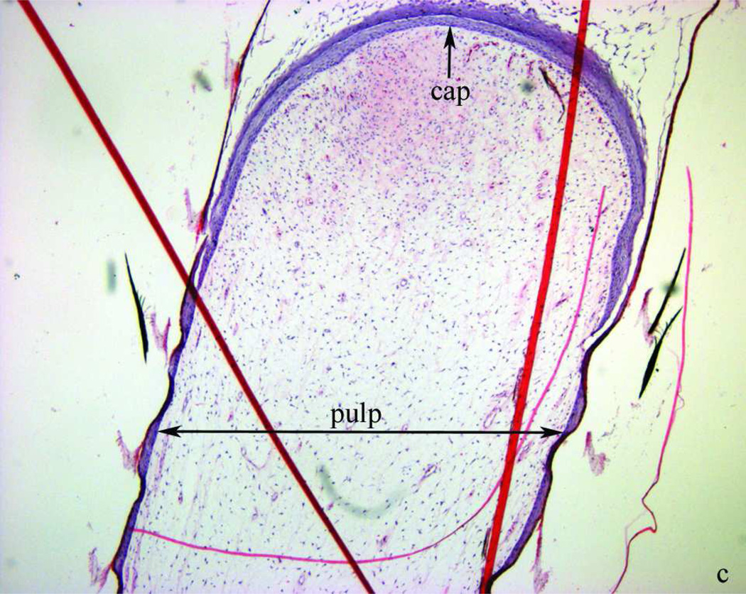
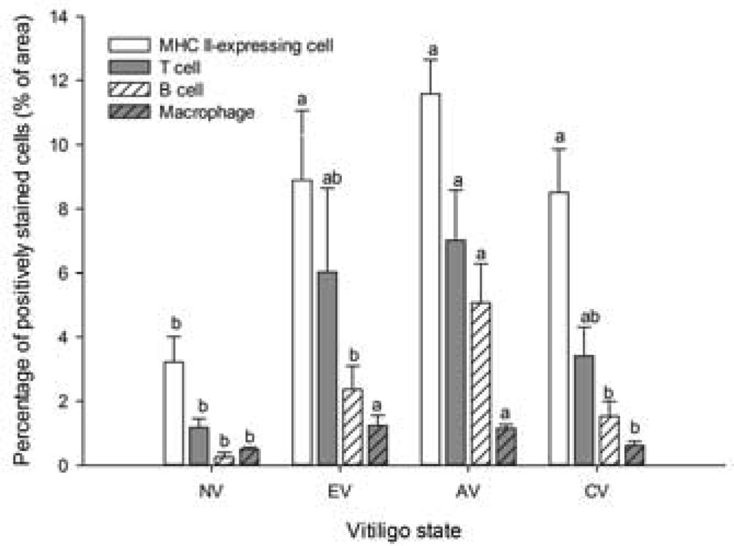
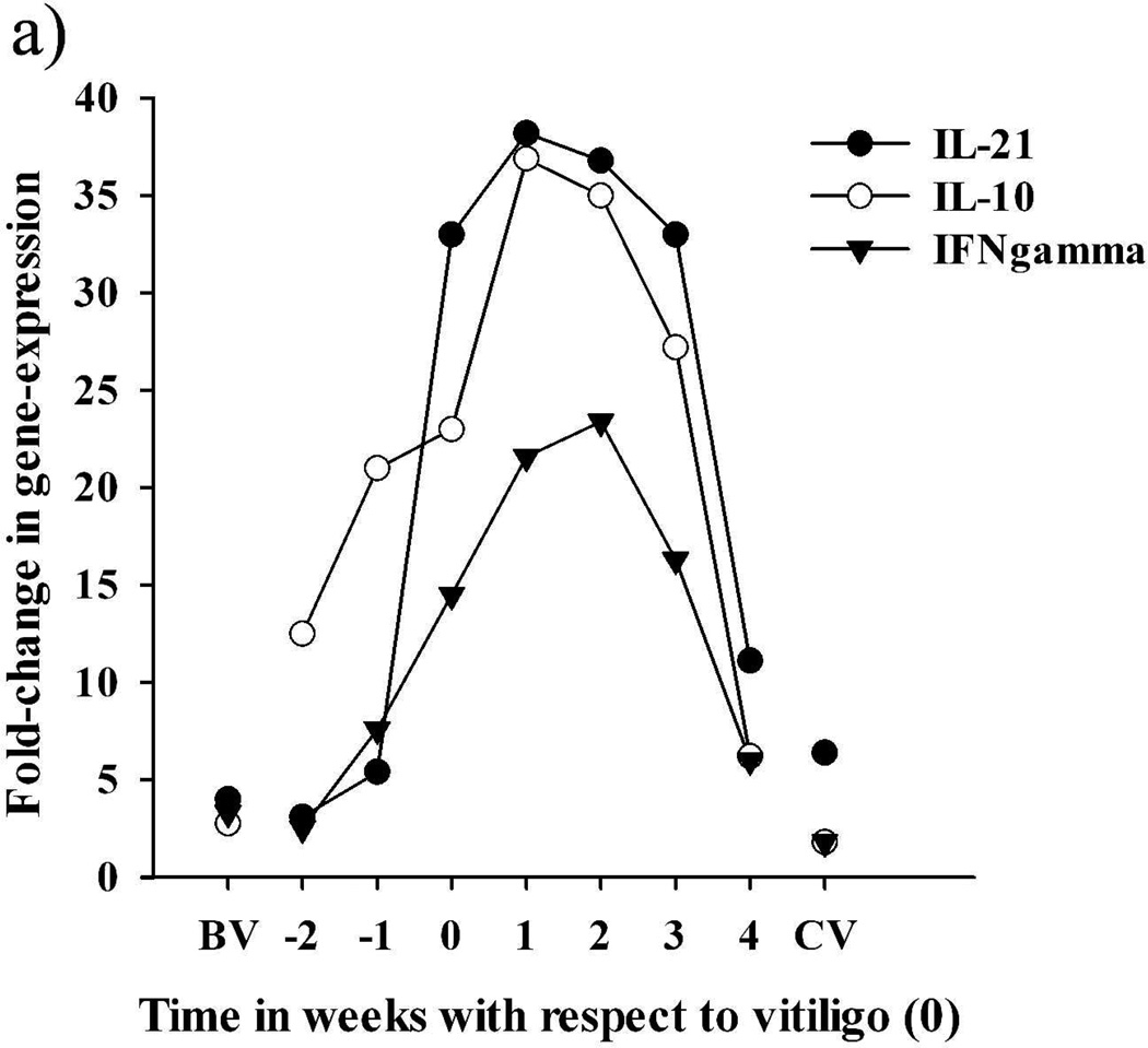
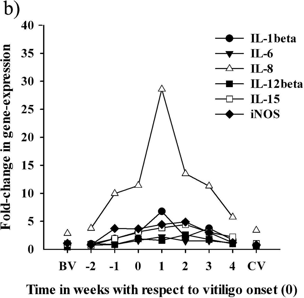
The Smyth line (SL) of chicken is an excellent animal model for human autoimmune vitiligo. In SL vitiligo (SLV), postnatal loss of melanocytes in feathers appears to be due to cell-mediated immunity. In this study, leukocyte infiltration and associated expression (RNA) of immune function-related cytokines in growing feathers were investigated throughout SLV development and progression. Both leukocyte infiltration and cytokine expression levels started to increase near visible SLV onset (early SLV), reached peak levels during active SLV, and decreased to near pre-vitiligo levels after complete loss of melanocytes. Specifically, significant increases were noticed in relative proportions of T cells, B cells, and major histocompatibility complex (MHC) II-expressing cells during active SLV. Levels of T-cell infiltration were higher than those of B cells, with more CD8+ than CD4+ cells throughout SLV. Elevated leukocyte infiltration in early and active SLV was accompanied by increased levels of cytokine expression, especially in IFN-γ, IL-10, and IL-21. Low expression of IL-4 and IL-17 did not suggest important roles of Th2 and Th17 cells in SLV pathogenesis. Taken together, SLV appears to be a Th1-polarized autoimmune disease, whereby IFN-γ expression is strongly associated with parallel increases in IL-10 and IL-21, particularly during early and active stages of SLV.


Conflict of interest statement

Competing interests

The authors state no conflict of interest.

Figures

Figure 1
Figure 1
Morphology of two- to three-week-old growing feathers from SL chickens. a) from left to right: normally pigmented, partially depigmented and completely depigmented growing feathers from SL chickens that developed SLV. Growing feathers can be collected from SLV chickens over the whole course of SLV. The living part of growing feathers (newest growth to the epidermal cap) is referred to as “feather tip”. b) microstructure of the newest growth of a feather tip with normal pigmentation; layers shown from the outside to inside are sheath, barb ridge and pulp. c) a cap formed by epidermal layer enclosing the pulp. Longitudinal sections were stained with H&E stain and examined at 40× (b, c) magnification under a bright field microscope. Bar scale = 1 mm.
Figure 1
Figure 1
Morphology of two- to three-week-old growing feathers from SL chickens. a) from left to right: normally pigmented, partially depigmented and completely depigmented growing feathers from SL chickens that developed SLV. Growing feathers can be collected from SLV chickens over the whole course of SLV. The living part of growing feathers (newest growth to the epidermal cap) is referred to as “feather tip”. b) microstructure of the newest growth of a feather tip with normal pigmentation; layers shown from the outside to inside are sheath, barb ridge and pulp. c) a cap formed by epidermal layer enclosing the pulp. Longitudinal sections were stained with H&E stain and examined at 40× (b, c) magnification under a bright field microscope. Bar scale = 1 mm.
Figure 2
Figure 2
Infiltration profiles of leukocytes in feather tips collected at different states of SLV. MHC II-expressing cells, T cells, B cells and macrophages were identified by indirect immunoperoxidase staining using Ia, TCR gamma-delta and TCR alpha-beta, Bu-1, and KUL01 mouse-anti-chicken monoclonal antibodies, respectively. Stained sections were examined at 40× magnification and the amount of stained cells expressed as the percentage of the whole feather section area analyzed (% of area). NV: feather tips from 3 SL chickens that never developed SLV; E V, AV and CV: feather tips were collected from 7 SLV chickens just before, during and after SLV development, respectively. Each bar represents the mean ± SE. a,b For each cell type, means without common letters are different (P < 0.05).
Figure 3
Figure 3
Time-course of the relative expression of cytokines and iNOS in growing feathers collected from seven SLV chickens throughout vitiligo development. Relative expression was calculated by the delta delta Ct method, using a cDNA pool made from growing feather of 3 SL chickens that never developed vitiligo as the calibrator and chicken 28S as the endogenous control gene. The X-axis represent the time (weeks) of feather tip collection with respect to vitiligo onset (0); BV and CV represent data from feathers collected from the 7 SL chickens >2 weeks before vitiligo and >1 week after complete pigmentation loss, respectively; weeks −2 and −1 represent early vitiligo (EV) and weeks 0–4 correspond to active vitiligo (AV). (a) Expression pattern of IL-21, IL-10, and IFN-gamma. (b) Expression pattern of IL-1beta, IL-6, IL-8, IL-12beta, IL-15 and iNOS. Also see Table 2.
Figure 3
Figure 3
Time-course of the relative expression of cytokines and iNOS in growing feathers collected from seven SLV chickens throughout vitiligo development. Relative expression was calculated by the delta delta Ct method, using a cDNA pool made from growing feather of 3 SL chickens that never developed vitiligo as the calibrator and chicken 28S as the endogenous control gene. The X-axis represent the time (weeks) of feather tip collection with respect to vitiligo onset (0); BV and CV represent data from feathers collected from the 7 SL chickens >2 weeks before vitiligo and >1 week after complete pigmentation loss, respectively; weeks −2 and −1 represent early vitiligo (EV) and weeks 0–4 correspond to active vitiligo (AV). (a) Expression pattern of IL-21, IL-10, and IFN-gamma. (b) Expression pattern of IL-1beta, IL-6, IL-8, IL-12beta, IL-15 and iNOS. Also see Table 2.

References

    1. Abbas A, Lichtman A, Pillai S, editors. Cellular and Molecular Immunology. Philadelphia: SaundersElsevier; 2010. p. 566.
    1. Abdul-Careem MF, Hunter DB, Shanmuganathan S, et al. Cellular and cytokine responses in feathers of chickens vaccinated against Marek's disease. Vet Immunol Immunopathol. 2008;126:362–366. - PubMed
    1. Austin LM, Boissy RE, Jacobson BS, et al. The detection of melanocyte autoantibodies in the Smyth chicken model for vitiligo. Clin Immunol Immunopathol. 1992;64:112–120. - PubMed
    1. Bassiouny DA, Shaker O. Role of interleukin-17 in the pathogenesis of vitiligo. Clin Exp Dermatol. 2011;36:292–297. - PubMed
    1. Boissy RE, Lamont SJ, Smyth JR., Jr. Persistence of abnormal melanocytes in immunosuppressed chickens of the autoimmune "DAM" line. Cell Tissue Res. 1984;235:663–668. - PubMed

Publication types

MeSH terms