. 2012 Feb;241(2):340-9.
doi: 10.1002/dvdy.22786. Epub 2011 Nov 23.

MCS9.7 enhancer activity is highly, but not completely, associated with expression of Irf6 and p63

Affiliations

MCS9.7 enhancer activity is highly, but not completely, associated with expression of Irf6 and p63

Walid D Fakhouri et al. Dev Dyn. 2012 Feb.

Abstract

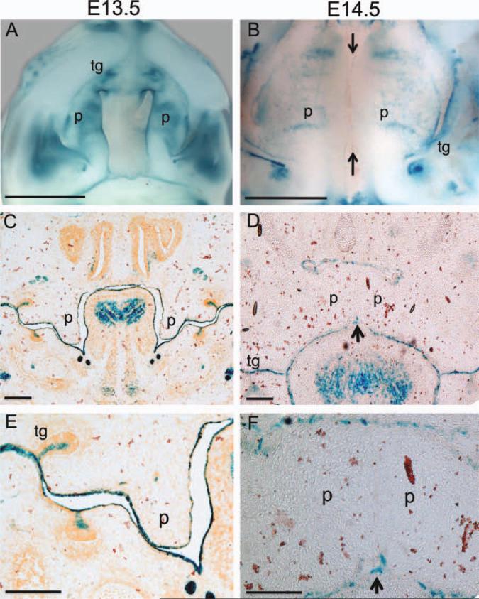
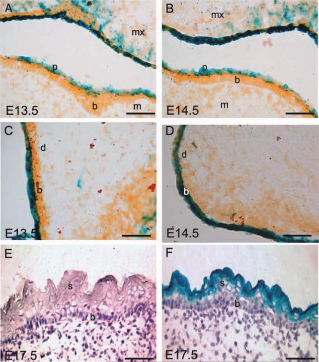
Background: DNA variation in Interferon Regulatory Factor 6 (IRF6) contributes risk for orofacial clefting, including a common DNA variant rs642961. This DNA variant is located in a multi-species conserved sequence that is 9.7 kb upstream from the IRF6 transcriptional start site (MCS9.7). The MCS9.7 element was shown to possess enhancer activity that mimicked the expression of endogenous Irf6 at embryonic day 11.5 in transient transgenic embryos, and also contains a p63 binding site that transactivates IRF6 expression. To analyze whether the MCS9.7 enhancer is sufficient to drive IRF6 expression, we generated stable transgenic murine lines that carry a MCS9.7-lacZ transgene. We hypothesized that MCS9.7 was sufficient to recapitulate the endogenous expression of Irf6 at other time-points during embryonic development.

Results: We observed that MCS9.7 activity recapitulated endogenous Irf6 expression in most tissues, but not in the medial edge epithelium (MEE) at E14.5, when Irf6 expression was high during secondary palatal fusion. Also, while MCS9.7 activity and Irf6 expression were associated with p63 expression, we observed MCS9.7 activity and Irf6 expression in periderm, although p63 was absent.

Conclusion: These data suggest that MCS9.7 enhancer activity is not sufficient to recapitulate IRF6 expression, and that p63 expression is not always necessary nor sufficient for transactivation of IRF6.


Figures

Fig. 1
Fig. 1
MCS9.7-lacZ enhancer activity during embryonic development. Bgal activity was visualized in whole-mount murine embryos between embryonic day (E) 9.5 and E13.5. A–D: Bgal activity was detected in the 1st (I), 2nd (II), and 3rd (III) branchial arches (A,B), maxillary (mx) and mandibular (m) processes (A,B), hindbrain (hb; A,B), dorsal surface of fore- (flb) and hindlimb buds (hlb; C,D), apical ectoderm ridge (aer; B), and the somites (s; A–D). Scale bar = 1.0 mm.
Fig. 2
Fig. 2
MCS9.7-lacZ activity in craniofacial tissues. Bgal activity was visualized in whole-mount transgenic murine embryos between embryonic day (E) 10.5 and E14.5. A–H: Frontal (A–D) and lateral (E–H) views of the same head. Bgal activity was observed in the medial nasal processes (mnp), maxillary processes (mx), mandibular processes (m), hindbrain (hb), nares (n), 2nd branchial arch (II), vibrissae (v), sub-mandibular gland (sg), and outer ear (e). Scale bar = 1.0 mm.
Fig. 3
Fig. 3
MCS9.7-lacZ enhancer activity in the oral cavity. A: Bgal activity in the medial edge and rugae of secondary palatal shelves (p) and in the incisor and molar tooth germs (tg) from whole-mount staining at embryonic day (E) 13.5. B: At E14.5, Bgal activity was similar except it was absent at the medial edge of the palatal shelves (arrows). C,E: Staining of coronal sections of E13.5 heads showed Bgal activity along oral, nasal, and tongue epithelium. D,F: However, Bgal activity was not observed in the medial edge epithelium of secondary palatal shelves just prior to palatal fusion despite the dark blue staining in oral epithelium and tooth germ. E and F images are close up for C and D. Scale bar = 0.75 mm (A,B), 0.5 mm (C,D), and 0.2 mm (E,F).
Fig. 4
Fig. 4
Expression of Krt14, Irf6, p63, and Bgal proteins in embryonic murine secondary palate. Fluorescent immunostaining was performed on serial coronal sections of heads of MCS9.7-lacZ transgenic embryo at embryonic day (E) 13.5 and E14.5. A–C: Krt14 serves as a marker for epithelial cells, including the medial edge epithelium (MEE). D–F: Irf6 is expressed in MEE throughout palatal development. G–I: Bgal was observed in all epithelium, including MEE at E13.5 (G), but was not detected in the MEE at E14.5 (H, I). J–L: Staining for p63 was identical to Bgal. Secondary palatal shelf (p); nasal septum (ns). White arrows indicate the MEE. Scale bar = 100 μm (A, B) and 50 μm (C).
Fig. 5
Fig. 5
Dynamic epithelium-specific enhancer activity of MCS9.7. A,B: In the oral epithelium, Bgal activity was observed primarily in the periderm at embryonic day (E) 13.5 (A) but was also observed in the basal layer at E14.5 (B). C,D: A similar change in expression was observed in the facial epidermis from E13.5, (C) to E14.5 (D). E,F: At E17.5, Bgal activity was not observed in transgene negative embryonic back skin (E), yet was detected in the suprabasal layers in transgene-positive animal (F). Maxilla (mx), mandible (m), basal layer (b), periderm (p), dermis (d), suprabasal layer (s). Scale bar = 200 μm (A-F).
Fig. 6
Fig. 6
Dynamic expression of Irf6 and p63 in palatal development. A,B: At embryonic day (E) 13.5 (A), expression of Irf6 (red) and p63 (green) appeared primarily distinct, but at E14.5 (B) they appeared to overlap primarily. Higher magnification showed p63 only in the basal layer at E13.5 (A1, A2), with Irf6 primarily in the periderm (A1) with some localized expression in basal cells (A2). At E14.5, both p63 and Irf6 were observed in the basal layer of oral epithelium (B1), but while p63 was greatly decreased in the medial edge epithelium (MEE), Irf6 remained strong (B2). Maxilla (mx), mandible (m), basal layer (b), dermis (d), palate (p). Scale bar = 100 μm (A, B) and 50 μm (A1, A2, B1, B2). A and B were stretched horizontally, and A was compressed vertically to make the images fit the figure.
Fig. 7
Fig. 7
A proposed model for the regulation of Irf6 in medial edge epithelium (MEE). Solid lines are based on our studies and previously published data (Thomason et al., 2010; Moretti et al., 2010). Dashed line represents a hypothetical regulation.

References

    1. Bailey CM, Khalkhali-Ellis Z, Kondo S, Margaryan NV, Seftor RE, Wheaton WW, Amir S, Pins MR, Schutte BC, Hendrix MJ. Mammary serine protease inhibitor (Maspin) binds directly to interferon regulatory factor 6: identification of a novel serpin partnership. J Biol Chem. 2005;280:34210–34217. - PMC - PubMed
    1. Beaty TH, Murray JC, Marazita ML, Munger RG, Ruczinski I, Hetmanski JB, Liang KY, Wu T, Murray T, Fallin MD, Redett RA, Raymond G, Schwender H, Jin SC, Cooper ME, Dunnwald M, Mansilla MA, Leslie E, Bullard S, Lidral AC, Moreno LM, Menezes R, Vieira AR, Petrin A, Wilcox AJ, Lie RT, Jabs EW, Wu-Chou YH, Chen PK, Wang H, Ye X, Huang S, Yeow V, Chong SS, Jee SH, Shi B, Christensen K, Melbye M, Doheny KF, Pugh EW, Ling H, Castilla EE, Czeizel AE, Ma L, Field LL, Brody L, Pangilinan F, Mills JL, Molloy AM, Kirke PN, Scott JM, Arcos-Burgos M, Scott AF. A genome-wide association study of cleft lip with and without cleft palate identifies risk variants near MAFB and ABCA4. Nat Genet. 2010;42:525–529. - PMC - PubMed
    1. Biggs LC, Rhea L, Schutte BC, Dunnwald M. Interferon regulatory factor 6 is necessary but not sufficient for keratinocyte differentiation. J Invest Derm. 2011 [Epub ahead of print] - PMC - PubMed
    1. Birnbaum S, Ludwig KU, Reutter H, Herms S, de Assis NA, Diaz-Lacava A, Barth S, Lauster C, Schmidt G, Scheer M, Saffar M, Martini M, Reich RH, Schiefke F, Hemprich A, Potzsch S, Potzsch B, Wienker TF, Hoffmann P, Knapp M, Kramer FJ, Nothen MM, Mangold E. IRF6 gene variants in Central European patients with nonsyndromic cleft lip with or without cleft palate. Eur J Oral Sci. 2009;117:766–769. - PubMed
    1. Burdick AB, Bixler D, Puckett CL. Genetic analysis in families with van der Woude syndrome. J Craniofac Genet Dev Biol. 1985;5:181–208. - PubMed

Publication types

LinkOut - more resources