. 2012 Feb;33(2):275-84.
doi: 10.1093/carcin/bgr276. Epub 2011 Nov 23.

Deletion of the SPARC acidic domain or EGF-like module reduces SPARC-induced migration and signaling through p38 MAPK/HSP27 in glioma

Affiliations

Deletion of the SPARC acidic domain or EGF-like module reduces SPARC-induced migration and signaling through p38 MAPK/HSP27 in glioma

Heather M McClung et al. Carcinogenesis. 2012 Feb.

Abstract

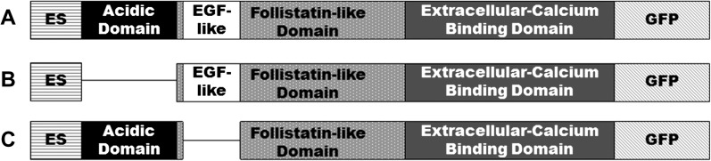
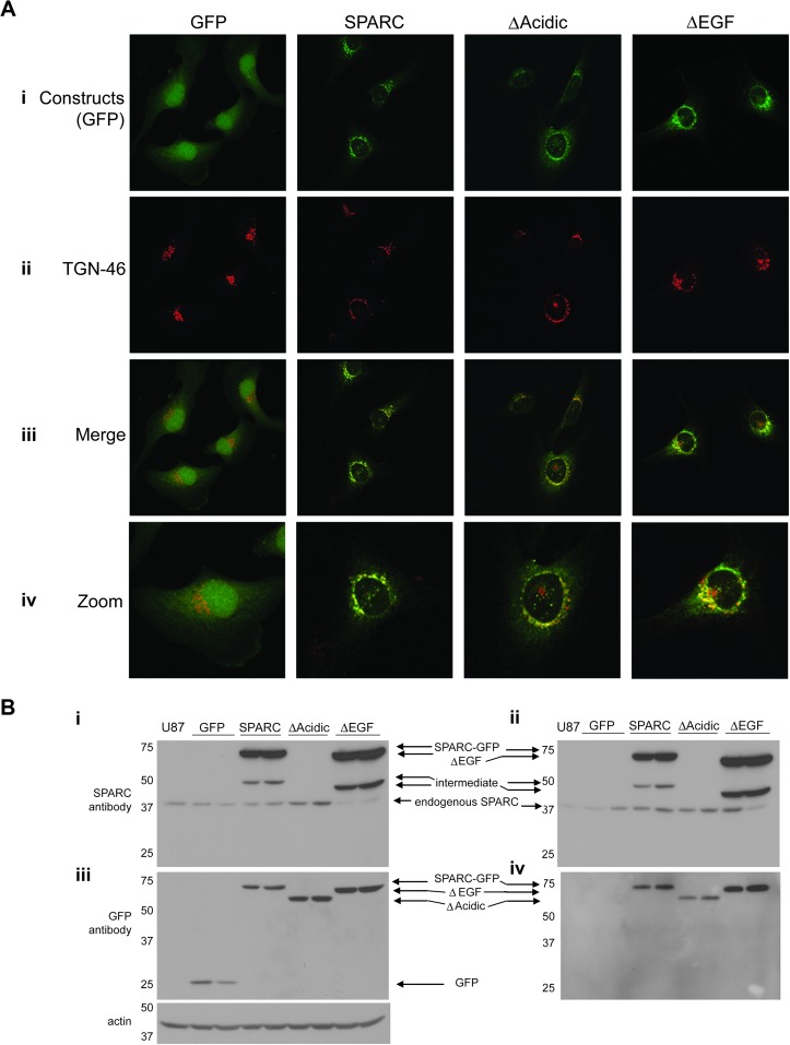
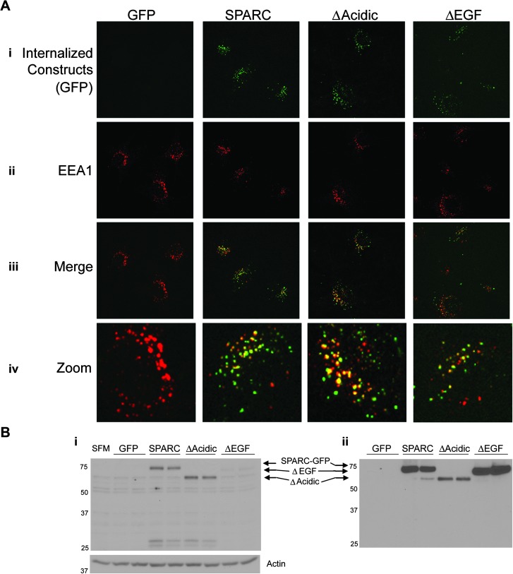
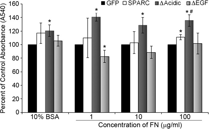
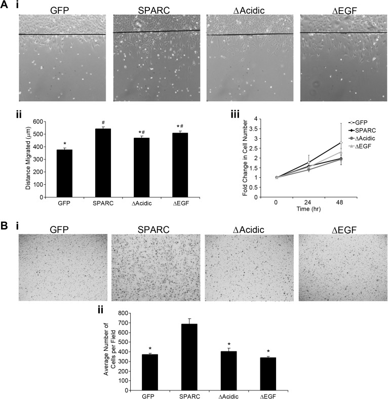
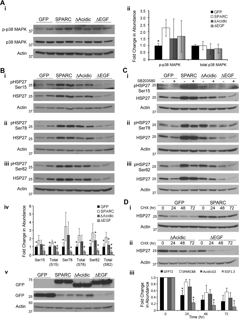
We previously demonstrated that secreted protein acidic and rich in cysteine (SPARC) increases heat shock protein 27 (HSP27) expression and phosphorylation and promotes glioma cell migration through the p38 mitogen-activated protein kinase (MAPK)/HSP27 signaling pathway. As different regions of the SPARC protein mediate different SPARC functions, elucidating which SPARC domains regulate HSP27 expression, signaling and migration might provide potential therapeutic strategies to target these functions. To investigate the roles of specific domains, we used an SPARC-green fluorescent protein (GFP) fusion protein and constructs of SPARC-GFP with deletions of either the acidic domain (ΔAcidic) or the epidermal growth factor (EGF)-like module (ΔEGF). GFP, SPARC-GFP and the two deletion mutants were expressed in U87MG glioma cells. Characterization of the derived stable clones by confocal imaging and western blotting suggests proper folding, processing and secretion of the deletion constructs. Uptake of the constructs by naive cells suggests enhanced internalization of ΔAcidic and reduced internalization of ΔEGF. Wound and transwell migration assays and western blot analysis confirm our previous results and indicate that ΔAcidic reduces SPARC-induced migration and p38 MAPK/HSP27 signaling and ΔEGF decreases SPARC-induced migration and dramatically decreases the expression and phosphorylation of HSP27 but is poorly internalized. Loss of the EGF-like module suppresses the enhanced HSP27 protein stability conferred by SPARC. In conclusion, deletions of the acidic domain and EGF-like module have differential effects on cell surface binding and HSP27 protein stability; however, both regions regulate SPARC-induced migration and signaling through HSP27. Our data link the domains of SPARC with different functions and suggest one or both of the constructs as potential therapeutic agents to inhibit SPARC-induced migration.


Figures

Fig. 1.
Fig. 1.
Schematic of the deletion constructs. (A) wt-SPARC–GFP contains the acidic domain (aa 1–52; black), follistatin-like domain (aa 53–130; gray with spots), which includes the EGF-like module (aa 55–76; white) and the β1 integrin-binding site (aa 113–130) and the extracellular calcium-binding domain (aa 131–280; solid gray). ES (horizontal stripes) indicates the export signal, which is not part of the mature protein, and GFP (diagonal stripes) refers to the C-terminal GFP tag. (B) The acidic domain deletion mutant construct has amino acids 1–52 of the mature protein deleted. (C) The EGF-like module deletion mutant construct has amino acids 55–76 deleted. aa, amino acids.
Fig. 2.
Fig. 2.
Expression and intracellular processing of the constructs. (A) Built confocal images showing one clone expressing each construct. (i) Green fluorescence shows that the control GFP is localized diffusely throughout the cells, whereas SPARC–GFP and the deletion mutants are localized perinuclearly. (ii) TGN-46 immunostaining indicates the trans-Golgi Network. (iii) Merged images indicate co-localization of TGN-46 with SPARC–GFP and both deletion mutants, confirming their localization to the Golgi complex. (iv) Zoomed images better demonstrate co-localization. Images in i–iii were captured at ×60. Zoomed images are ×240. (B) Western blot analyses of cell lysates (i and iii) and conditioned medium (ii and iv) demonstrate the level of intracellular and secreted constructs and endogenous wt-SPARC by the clones. The parental cell line, U87MG, is represented in lane 1 of each blot. (i and ii) Anti-SPARC antibody detects endogenous SPARC, SPARC–GFP and ΔEGF but not ΔAcidic or GFP. The intermediate bands are believed to be due to alternate processing or proteolytic cleavage. (iii and iv) Anti-GFP antibody detects all of the constructs (GFP is not secreted). Actin indicates equal loading of cell lysates. Molecular weights (in kDa) are shown at the left of each blot.
Fig. 3.
Fig. 3.
Internalization of the constructs by naive cells. (A) Built confocal images showing U87D8 exposed to conditioned medium from one clone per construct for 3 h. (i) Internalized constructs. Note that GFP is not secreted and so there is no GFP in the conditioned medium to be taken up by the cells. (ii) EEA1 immunostaining indicates the early endosome. (iii) Merged images show co-localization of EEA1 with SPARC–GFP, ΔAcidic and ΔEGF, indicating that they are internalized into the endosomes. Images i–iii were captured at ×60. (iv) Zoomed images (×240) better demonstrate co-localization. (B) Western blot analysis. (i) Lysates collected from U87D8 after 6 h exposure to conditioned media or SF OptiMEM (SFM) shows that SPARC–GFP and both deletion mutants are present in cell lysates of the naive cells. The blot is labeled according to the conditioned media to which the U87D8 cells were exposed. Actin indicates equal loading of cell lysates. (ii) The level of SPARC–GFP or deletion mutant constructs present in the conditioned media. Molecular weights (in kDa) are indicated at the left of each blot.
Fig. 4.
Fig. 4.
Deletion of the acidic domain increases cell adhesion. Adhesion was measured at 24 h. SPARC-expressing cells were more adherent than GFP cells on the highest level of FN. Expression of ΔAcidic increased adhesion compared with control on all levels of FN and on BSA. ΔAcidic increased adhesion on 100 μg/ml FN compared with SPARC. Expression of ΔEGF decreased adhesion compared with control cells on 1 μg/ml FN. ΔEGF cells were not significantly different from SPARC but were less adherent than ΔAcidic on all concentrations of FN (P ≤ 0.03). Concentration of FN shown is in μg/ml. * Significantly different from GFP (P ≤ 0.036), #Significantly greater than SPARC (P = 0.0095).
Fig. 5.
Fig. 5.
Deletion of the acidic domain or the EGF-like module reduces SPARC-induced migration. (A) Wound migration assay. (i) Representative ×10 images of one clone expressing each construct indicate cell migration from the start of the wound after 20 h. (ii) Average distance migrated for both clones expressing each construct. SPARC significantly increased migration over control. Deletion of the acidic domain or the EGF-like module reduced migration compared with SPARC. The deletion mutants migrated farther than control cells. (iii) Fold change in cell number at 24 and 48 h relative to 0 h indicates that increased migration is not due to increased proliferation. (B) Transwell migration assay. (i) Representative ×10 images of one clone expressing each construct indicate migration through transwell filters after 2 h. (ii) Average number of cells per field. Expression of SPARC–GFP increases cell migration compared with control cells. Both deletions reduced migration to control levels. *Significantly less than SPARC (P ≤ 0.033), #Significantly greater than GFP (P < 0.01).
Fig. 6.
Fig. 6.
Deletion of the acidic domain or EGF-like module reduces SPARC-mediated signaling through the p38 MAPK/HSP27 pathway. (A) (i) Western blot analysis of expression and activation of p38 MAPK and (ii) densitometric analysis indicate that SPARC increases phosphorylation of p38 MAPK. The deletion mutants were not significantly different from GFP- or SPARC-expressing cells. (B) Western blot analysis of HSP27 expression and phosphorylation at (i) serine 15, (ii) serine 78 and (iii) serine 82 and (iv) densitometric analysis show that SPARC increases HSP27 expression and phosphorylation at all three serines. Deletion of the acidic domain reduces HSP27 expression and phosphorylation at serine 82. ΔAcidic was not significantly different from control cells. Deletion of the EGF-like module decreased HSP27 expression and phosphorylation to or below control levels. Equal expression of the constructs is confirmed using anti-GFP antibody in v. (C) Cells were treated with 20 μM SB203580 for 24 h. Western blot analysis of HSP27 expression and phosphorylation at (i) serine 15, (ii) serine 78 and (iii) serine 82 indicates that HSP27 phosphorylation is mediated by p38 MAPK. (D) Cells were exposed to 50 μg/ml cycloheximide for the times indicated. (i, ii) Western blot analysis and (iii) densitometric analysis indicate that the increased levels of HSP27 in the SPARC-expressing clones is due at least in part to increased protein stability and this increased stability is lost with the deletion of the EGF-like module. * Significantly less than SPARC (P < 0.05), #Significantly less than GFP (P < 0.016). Molecular weights (in kDa) are indicated at the left of each blot.

References

    1. Rempel SA, et al. Tumor invasiveness and anti-invasion strategies. In: Newton HB, editor. Handbook of Brain Tumor Chemotherapy. 2006. Elsevier Inc., San Diego, CA, pp. 193–218.
    1. Lefranc F, et al. Possible future issues in the treatment of glioblastomas: special emphasis on cell migration and the resistance of migrating glioblastoma cells to apoptosis. J. Clin. Oncol. 2005;23:2411–2422. - PubMed
    1. Louis DN. Molecular pathology of malignant gliomas. Annu. Rev. Pathol. 2006;1:97–117. - PubMed
    1. Demuth T, et al. Molecular mechanisms of glioma cell migration and invasion. J. Neurooncol. 2004;70:217–228. - PubMed
    1. Sage H, et al. Characterization of a novel serum albumin-binding glycoprotein secreted by endothelial cells in culture. J. Biol. Chem. 1984;259:3993–4007. - PubMed

Publication types

MeSH terms