. 2012 Feb 1;302(3):L352-9.
doi: 10.1152/ajplung.00302.2011. Epub 2011 Nov 23.

Hypoxia-inducible factor-1α regulates KCNMB1 expression in human pulmonary artery smooth muscle cells

Affiliations

Hypoxia-inducible factor-1α regulates KCNMB1 expression in human pulmonary artery smooth muscle cells

Yong-Tae Ahn et al. Am J Physiol Lung Cell Mol Physiol. .

Abstract

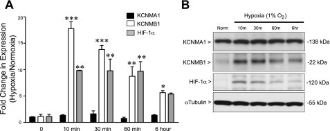
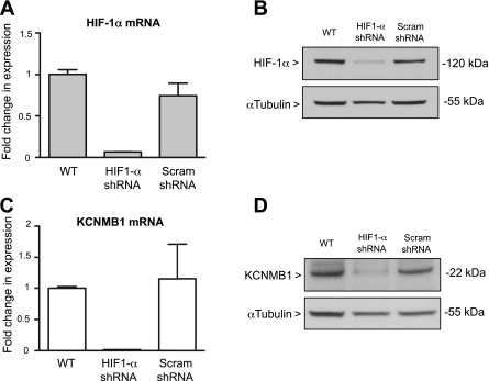
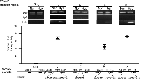
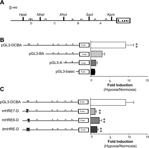
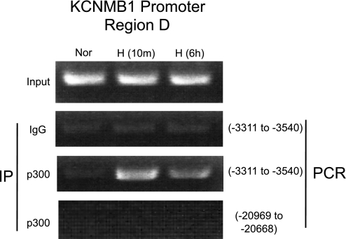
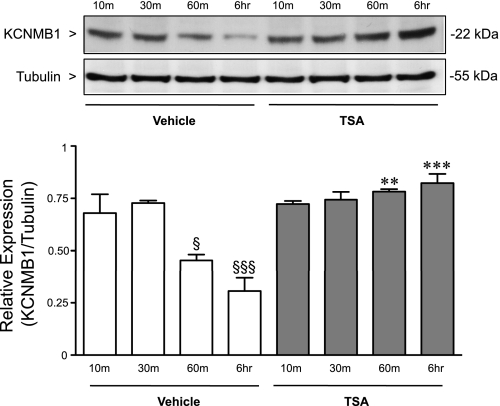
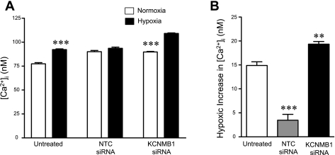
Previously, we observed that hypoxia increases the expression of the β1-subunit (KCNMB1) of the calcium-sensitive potassium channel (BK(Ca)). Herein, we elucidate the mechanism whereby hypoxia increases KCNMB1 expression in human pulmonary artery smooth muscle cells (hPASMC). In response to hypoxia, the expression of both the transcription factor hypoxia-inducible factor 1-α (HIF-1α) and KCNMB1 are increased. Knockdown of HIF-1α using a shRNA plasmid blocked the hypoxic induction of KCNMB1 expression. Chromatin immunoprecipitation (ChIP) demonstrated HIF-1α binding to three discrete regions of the human KCNMB1 promoter known to contain hypoxia response elements (HREs). A KCNMB1 promoter reporter assay combined with site-directed mutagenesis identified two adjacent HREs located between -3,540 bp and -3,311 bp that are essential for the hypoxic induction of KCNMB1 promoter activity. Furthermore, additional ChIP assays demonstrated recruitment of the HIF-1α transcriptional coactivator, p300, to this same promoter region. Treatment of hPASMC with the histone deacetylase inhibitor, trichostatin, prolonged the increase in KCNMB1 observed with hypoxia, suggesting that alterations in chromatin remodeling function to limit the hypoxic induction of KCNMB1. Finally, KCNMB1 knockdown potentiated the hypoxia-induced increase in cytosolic calcium in hPASMC, highlighting the contribution of the β1-subunit in modulating vascular SMC tone in response to acute hypoxia. In conclusion, HIF-1α increases KCNMB1 expression in response to hypoxia in hPASMC by binding to two HREs located at -3,540 to -3,311 of the KCNMB1 promoter. We speculate that selective modulation of KCNMB1 expression may serve as a novel therapeutic approach to address diseases characterized by an increase in vascular tone.


Figures

Fig. 1.
Fig. 1.
Hypoxia increases the β1-subunit (KCNMB1) of the calcium-sensitive potassium channel (BKCa) and hypoxia-inducible factor-1α (HIF-1α) in human pulmonary artery smooth muscle cells (hPASMC). A: qRT-PCR in hPASMC demonstrates a significant increase in the α1-subunit of BKCa (KCNMB1) and HIF-1α mRNA expression after 10 min to 6 h of hypoxia compared with normoxia. No difference was found in KCNMA1 gene expression. *P < 0.05, **P < 0.01, and ***P < 0.001 vs. normoxia by one-way ANOVA followed by Bonferroni posttest analysis, with n = 3. B: representative Western immunoblot to detect KCNMA1, KCNMB1, and HIF-1α protein expression in hPASMC demonstrates that hypoxia increases KCNMB1 protein within 10 min of hypoxic exposure and persists through 6 h, which mirrors the increase in HIF-1α protein expression.
Fig. 2.
Fig. 2.
Silencing HIF-1α prevents the hypoxic induction of KCNMB1. A and B: qRT-PCR and Western immunoblot demonstrate that stable transfection of hPASMC with shRNA to target HIF-1α effectively decreases HIF-1α gene and protein expression. C and D: qRT-PCR and Western immunoblot to detect KCNMB1 mRNA and protein show that knockdown of HIF-1α prevents the hypoxic induction of KCNMB1 expression. In Western immunoblotting experiments, α-tubulin was used as a protein loading control. WT, wild-type; Scram; scrambled.
Fig. 3.
Fig. 3.
Hypoxia increases binding of HIF-1α to discrete regions of the human KCNMB1 promoter. Chromatin immunoprecipitation (ChIP) demonstrated that hypoxia induced binding of HIF-1α-containing complexes and the KCNMB1 promoter regions A, B, and D. Data are presented as relative binding activity determined as the intensity of the PCR product obtained after HIF-1α IP relative to the input signal and expressed as a percent, with n = 3. HRE, hypoxia response element.
Fig. 4.
Fig. 4.
HIF-1α-mediated transcription of KCNMB1 requires binding to the HREs located between −3,540 and −3,311 bp of the promoter. A: schematic of the KCNMB1 promoter divided into fragments arbitrarily named (A, B, C, and D) based on the distribution of HREs (gray boxes) and location of restriction enzyme sites. B: hPASMC were transfected with 1 of 3 luciferase reporter plasmids containing different fragments of the KCNMB1 promoter, or the negative control plasmid, pGL3-basic. Transfected hPASMCs were then exposed to 1% O2 for 6 h, and luciferase reporter activity was measured and reported as the fold induction of reporter activity after hypoxia vs. normoxia. **P < 0.01 vs. pGL3-basic, with data presented as the means ± SE of 3 independent experiments. C: hPASMC transfected with luciferase reporter constructs containing mutations within each of the 2 HRE located within the D region (mHRE7-D and mHRE6-D), or a double mutant with both HRE mutated (dmHRE-D). Transfected cells were exposed to 1% O2 for 6 h, and luciferase reporter activity was measured and reported as the fold induction of reporter activity after hypoxia vs. normoxia. **P < 0.01 vs. pGL3-DCBA, with data presented as the means ± SE of 3 independent experiments.
Fig. 5.
Fig. 5.
Hypoxia increases binding of the transcriptional coactivator, p300, to the KCNMB1 promoter at 3,540 to −3,311 bp. Representative ChIP after IP with an antibody against p300 followed by and PCR using primers designed to detect the D region (−3,540 to −3,311 bp) of the KCNMB1 promoter in hPASMC under normoxia, and after either 10 min or 6 h of hypoxia. Control experiments were performed by immunoprecipitating with an isotype control antibody (Ig), and after IP using the p300 antibody followed by PCR using primers corresponding to an upstream region of the KCNMB1 promoter that does not contain an HRE.
Fig. 6.
Fig. 6.
Histone deacetylase (HDAC) inhibition prolongs the hypoxic induction of KCNMB1. Western immunoblot to detect KCNMB1 protein after treatment of the hPASMC with either vehicle, or the HDAC inhibitor, trichostatin A (TSA), and exposure to hypoxia for 10 min to 6 h. α-Tubulin was used as a loading control. §P < 0.05 vs. vehicle 30 min, and §§§P < 0.001 vs. vehicle 10 min and 30 min. **P < 0.01 vs. vehicle 60 min, and ***P < 0.001 vs. vehicle 6 h.
Fig. 7.
Fig. 7.
Depletion of KCNMB1 potentiates the response of hPASMC to hypoxia. hPASMC were transfected with either nontargeting (control) or KCNMB1 siRNA. Untreated and siRNA-transfected cells were loaded with fura-2 AM and exposed to acute hypoxia for 20 min. A: hypoxia caused a significant increase in cytosolic calcium (P < 0.05 vs. normoxia; n = 14). Transfection of hPASMC with nontargeted control siRNA blunted hypoxia-induced increase in of cytosolic calcium (n = 15). However, KCNMB1 knockdown with siRNA accentuated the response of hPASMC to hypoxia, resulting in significant increase in cytosolic calcium (***P < 0.001, vs. normoxic control for that treatment group, with n = 14–15, per group). B: after 20 min of hypoxia, untreated hPASMC demonstrated a significant increase in cytosolic calcium. Compared with untreated cells, the response to hypoxia in hPASMC treated with nontargeting (control) siRNA was attenuated (***P < 0.001). In hPASMC treated with KCNMB1 siRNA, the response to hypoxia was accentuated, compared with untreated cells (**P < 0.01). NTC, non targeted control.

References

    1. Amberg GC, Santana LF. Downregulation of the BK channel beta1 subunit in genetic hypertension. Circ Res 93: 965–971, 2003 - PubMed
    1. Brayden JE, Nelson MT. Regulation of arterial tone by activation of calcium-dependent potassium channels. Science 256: 534–535, 1992 - PubMed
    1. Brenner R, Peréz GJ, Bonev AD, Eckman DM, Kosek JC, Wiler SW, Patterson AJ, Nelson MT, Aldrich RW. Vasoregulation by the beta1 subunit of the calcium-activated potassium channel. Nature 407: 870–876, 2000 - PubMed
    1. Cornfield DN, Reeve HL, Tolarova S, Weir EK, Archer SL. Oxygen causes fetal pulmonary vasodilation through activation of a calcium-dependent potassium channel. Proc Natl Acad Sci USA 93: 8089–8094, 1996 - PMC - PubMed
    1. Cornfield DN, Saqueton CB, Porter VA, Herron JM, Resnik ER, Haddad IY, Reeve HL. Voltage-gated K+ channel activity in the ovine pulmonary vasculature is developmentally regulated. Am J Physiol Lung Cell Mol Physiol 278: L1297–L1304, 2000 - PubMed

Publication types

MeSH terms

Substances

LinkOut - more resources