. 2012 Feb;23(2):225-35.
doi: 10.1681/ASN.2011040429. Epub 2011 Nov 23.

Pathologic characteristics of transplanted kidney xenografts

Affiliations

Pathologic characteristics of transplanted kidney xenografts

Akira Shimizu et al. J Am Soc Nephrol. 2012 Feb.

Abstract

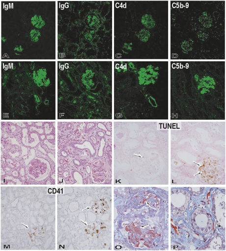
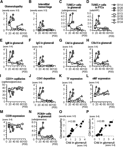
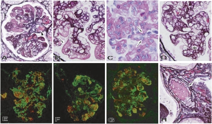
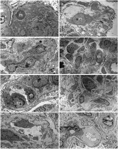
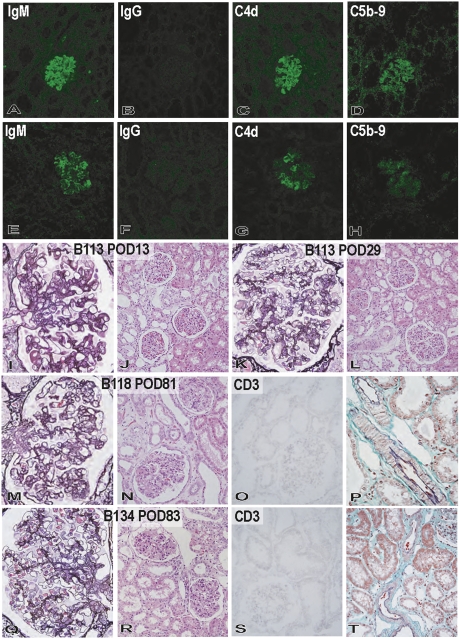
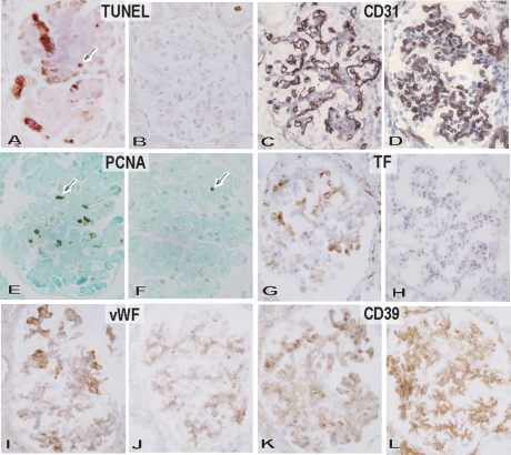
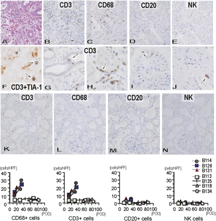
For xenotransplantation to become a clinical reality, we need to better understand the mechanisms of graft rejection or acceptance. We examined pathologic changes in α1,3-galactosyltransferase gene-knockout pig kidneys transplanted into baboons that were treated with a protocol designed to induce immunotolerance through thymic transplantation (n=4) or were treated with long-term immunosuppressants (n=3). Hyperacute rejection did not occur in α1,3-galactosyltransferase gene-knockout kidney xenografts. By 34 days, acute humoral rejection led to xenograft loss in all three xenografts in the long-term immunosuppression group. The failing grafts exhibited thrombotic microangiopathic glomerulopathy with multiple platelet-fibrin microthrombi, focal interstitial hemorrhage, and acute cellular xenograft rejection. Damaged glomeruli showed IgM, IgG, C4d, and C5b-9 deposition. They also demonstrated endothelial cell death, diffuse endothelial procoagulant activation with high expression of tissue factor and vWF, and low expression of the ectonucleotidase CD39. In contrast, in the immunotolerance group, two of four grafts had normal graft function and no pathologic findings of acute or chronic rejection at 56 and 83 days. One of the remaining kidneys had mild but transient graft dysfunction with reversible, mild microangiopathic glomerulopathy, probably associated with preformed antibodies. The other kidney in the immunotolerance group developed unstable graft function at 81 days and developed chronic xenograft glomerulopathy. In summary, the success of pig-to-primate xenotransplantation may necessitate immune tolerance to inhibit acute humoral and cellular xenograft rejection.


Figures

Figure 1.
Figure 1.
Pathologic features of the grafts during progression of graft failure in the immunosuppression protocol. Immunofluorescence studies of IgM (A and E: original magnification, ×200), IgG (B and F: original magnification, ×200), C4d (C and G: original magnification, ×200), and C5b-9 (D and H: original magnification, ×200) in the grafts (B114) at postoperative days 12 (A–D) and 33 (E–H) showed progression of immunoglobulin and complement deposition in glomeruli and small arteries and focally along peritubular capillaries. Development of graft failure, from postoperative days 12 (I, K, and M) to 33 (J, L, N–P), was associated with the development of thrombotic microangiopathic glomerulopathy (I and J: hematoxylin and eosin stain; original magnification, ×200) with TUNEL+ dead cells (arrow) (K and L: TUNEL stain, original magnification, ×200), CD41+ activated platelets (arrow) (M and N: CD41 stain; original magnification, ×200), and fibrin exudation (arrow) (O: Masson stain; original magnification, ×600). Interstitial edema and focal hemorrhage (J) and fibrin thrombi (arrowhead) in peritubular capillaries (P: Masson stain; original magnification, ×600) were also evident in the interstitium at postoperative day 33.
Figure 2.
Figure 2.
Pathologic differences between immunosuppression (B114, B126, and B131) and immunotolerance (B113, B135, B118, and B134) protocols. The pathologic characteristics were evaluated semiquantitatively for the severity of glomerulopathy (A); the degree of interstitial hemorrhage (B); the number of TUNEL+ dead cells in glomeruli (C) and peritubular capillaries (PTCs) (D); the glomerular deposition of IgM (E), IgG (F), C4d (G), and C5b-9 (H); the number of CD31+ glomerular capillaries (I); the deposition of CD41 platelets (J); the expression of tissue factor (K), vWF (L), and CD39 (M); and the number of PCNA+ proliferating glomerular cells (N). Correlation between C4d deposition in glomeruli and the severity of glomerulopathy (O) or CD41 deposition (P) was also evaluated. POD, postoperative day.
Figure 3.
Figure 3.
Development of thrombotic microangiopathic glomerulopathy and arteriolopathy in the immunosuppression protocol (A, F, and G: B114, postoperative day 33; B and H: B131, postoperative day 20; C and D: B126, postoperative day 34). Thrombotic microangiopathic glomerulopathy developed with multiple thrombi (arrowhead in A: periodic acid-methenamine [PAM] stain; original magnification, ×600), fibrin exudation (arrow in B: PAM stain; original magnification, ×800), enlarged endothelial cells (arrow in C: PAS stain; original magnification, ×800) and mesangial proliferative lesion (asterisk in D: PAM stain; original magnification, ×800). Almost all CD41+ platelet-rich thrombi were detected with fibrin; within vessels they were detected with deposition of IgM and C4d (double immunostaining against CD41 [red] and green of fibrin [E], IgM [F] or C4d [G]; original magnification, ×600). Thrombosis (arrow) was also evident in small arteries (H: PAM stain; original magnification, ×800).
Figure 4.
Figure 4.
Electron microscopic findings of xenografts of the immunosuppression and immunotolerance protocols. In thrombotic microangiopathic glomerulopathy at postoperative day 33 in B114 in the immunosuppression protocol, the glomerular capillary structure was lost with apoptotic cell (asterisk) and fibrin deposition (A: original magnification, ×5000), mesangial matrix-lysis and activated and enlarged mesangial cells (asterisk) (B: original magnification, ×4000), and activated and enlarged endothelial cells (asterisk) (C: original magnification, ×6000). In reversible microangiopathic glomerulopathy in the immunotolerant baboon (B113), swelling of endothelial cells (asterisk) was noted in glomeruli with partial dilatation of subendothelial space and mesangiolytic changes at postoperative day 13 (D: original magnification, ×2000; E; original magnification, ×7000). However, in the immunotolerance group, these findings recovered to almost intact glomerular structure by postoperative day 29 (B113: F; original magnification, ×2000). The glomeruli in the graft with unstable graft function from postoperative day 50 showed double contour of glomerular basement membrane (arrow in G), mesangial proliferation, and effacement of foot processes of podocytes at postoperative day 81 (B118: G; original magnification, ×7000). The glomeruli in the graft with stable graft function showed almost intact glomerular capillaries with preservation of foot process of podocytes at postoperative day 83 (B134: H; original magnification, ×5000).
Figure 5.
Figure 5.
Pathologic changes in the grafts of the immunotolerance protocol. In reversible glomerular injury in B113, deposition of IgM (A), C4d (C), and C5b-9 (D), but not IgG (B), occurred by postoperative day 13 (A–D: original magnification, ×200), and mild thrombotic microangiopathic glomerulopathy developed (I: periodic acid-methenamine [PAM] stain; original magnification, ×600). Cellular rejection and arteriolar changes were not detected at postoperative day 13 in B113 (J: hematoxylin and eosin stain; original magnification, ×200). However, these glomerular alterations recovered to almost their structure by postoperative day 29 in B113 (K: PAM stain; original magnification, ×600). Cellular rejection, arteriolar changes, and interstitial fibrosis did not develop at postoperative day 29 in B113 (L: hematoxylin and eosin stain; original magnification, ×200). In the graft with unstable graft function (B118), focal mesangial proliferation with double contour of glomerular basement membrane were seen at postoperative day 81 (M: PAM stain; original magnification, ×600). Focal interstitial fibrosis was noted, but cellular rejection and arterial changes were not detected (N: hematoxylin and eosin stain; original magnification, ×200; O: CD3 stain; original magnification, ×200; P: elastica-Masson Goldner stain; original magnification, ×600). In the graft with stable graft function (B134) at postoperative day 83, focal and weak deposition of IgM (E), C4d (G), and C5b-9 (H) was seen, but no deposition of IgG (F) was noted (E–H: original magnification, ×200). The glomeruli showed no obvious glomerulopathy at postoperative day 83 in B134 (Q: PAM stain; original magnification, ×600) with no cellular rejection or vascular changes (R: hematoxylin and eosin stain; original magnification, ×200; S: CD3 stain; original magnification, ×200; T: elastica-Masson Goldner stain; original magnification, ×200). POD, postoperative day.
Figure 6.
Figure 6.
Immunopathological characterization of thrombotic microangiopathic glomerulopathy. TUNEL+ dead cells (A and B: original magnification, ×600), CD31+ glomerular capillaries (C and D: original magnification, ×600), PCNA+ proliferating cells (E and F: original magnification, ×600), tissue factor expression (G and H: original magnification, ×600), vWF expression (I and J: original magnification, ×600), and CD39 expression (K and L: original magnification, ×600) in glomeruli of the immunosuppression (A, C, E, G, I, and K: B114, postoperative day 34) and immunotolerance (B, D, F, H, J, and L: B134, postoperative day 83) baboons. In the immunosuppression protocol, thrombotic microangiopathic glomerulopathy developed at postoperative day 34 in B114 with many TUNEL+ dead cells (arrow) (A), loss of CD31+ glomerular capillaries (C), proliferating PCNA+ cells (arrow) (E), increased expression of tissue factor (G) and vWF (I), and decreased expression of CD39 (K). In contrast, the glomeruli of the immunotolerance baboon (B134) at postoperative day 83 were characterized by preservation of CD31+ glomerular capillaries (D) with rare TUNEL+ dead cells (B) and PCNA+ cells (F), no upregulation of tissue factor (H) and vWF expression, and persistent CD39 expression (L).
Figure 7.
Figure 7.
Cellular infiltration within Gal knockout kidneys in baboons treated with chronic immunosuppression or immunotolerance protocol. Cellular infiltration in immunosuppression (A–J: B114, postoperative day 34) and immunotolerance (K–N: B134, postoperative day 83) protocols. Cellular infiltration developed at postoperative day 34 (B114) (A: hematoxylin and eosin stain; original magnification, ×400) in the immunosuppression protocol with infiltration of CD3+ T cells (B: original magnification, ×400) and CD68+ macrophages (C: original magnification, ×400), although with little infiltration of CD20+ B cells (D: original magnification, ×400) and NK cells (E: original magnification, ×400). Many CD3+ (brown) T cells contained TIA-1+ (blue) cytotoxic granules (arrow) (F: original magnification, ×900), capillaritis, acute glomerulitis, and endothelialitis developed with CD3+ T cells (arrow) in tubules (G: original magnification, ×800), peritubular capillaries (H: original magnification, ×800), glomeruli (I: original magnification, ×800), and arteries (J: original magnification, ×800). In contrast, only small numbers of CD3+ T cells (K: original magnification, ×200) and CD68+ macrophages (L: original magnification, ×200), and very few CD20+ B cells (M: original magnification, ×200) and NK cells (N: original magnification, ×200) were present in the grafts at postoperative day 83 in B134 in the immunotolerance protocol. Analysis of the number of infiltrating CD3+ T cells, macrophages, CD20+ B cells, and NK cells in the grafts (cells/single field of 0.0625 mm2) indicated increased infiltration of T cells and macrophages in the grafts of the immunosuppression protocol, but not those of the immunotolerance protocol. HPF, high-power field; POD, postoperative day.

Similar articles

Cited by

References

    1. Sachs DH, Sykes M, Robson SC, Cooper DK: Xenotransplantation. Adv Immunol 79: 129–223, 2001 - PubMed
    1. Cascalho M, Ogle BM, Platt JL: Xenotransplantation and the future of renal replacement. J Am Soc Nephrol 15: 1106–1112, 2004 - PubMed
    1. Cooper DK, Gollackner B, Sachs DH: Will the pig solve the transplantation backlog? Annu Rev Med 53: 133–147, 2002 - PubMed
    1. Sachs DH: The pig as a potential xenograft donor. Vet Immunol Immunopathol 43: 185–191, 1994 - PubMed
    1. Bach FH, Robson SC, Winkler H, Ferran C, Stuhlmeier KM, Wrighton CJ, Hancock WW: Barriers to xenotransplantation. Nat Med 1: 869–873, 1995 - PubMed

Publication types

Substances