

Acute renal venous obstruction is more detrimental to the kidney than arterial occlusion: implication for murine models of acute kidney injury
- PMID: 22114209
- PMCID: PMC3353642
- DOI: 10.1152/ajprenal.00011.2011
Acute renal venous obstruction is more detrimental to the kidney than arterial occlusion: implication for murine models of acute kidney injury
Abstract
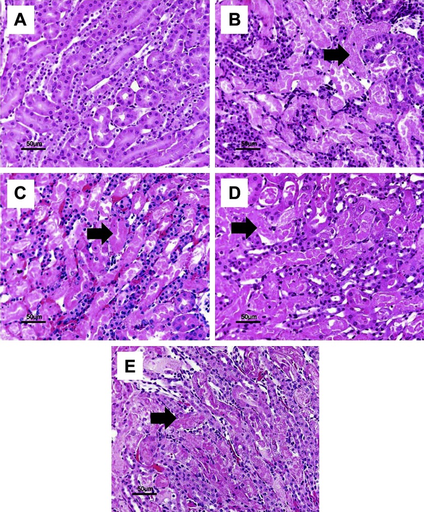
In this study, we compared the traditional murine model with renal pedicle clamp with models that clamped the renal artery or vein alone as well as to a whole body ischemia-reperfusion injury (WBIRI) model. Male C57BL/6J mice underwent either clamping of the renal artery, vein, or both (whole pedicle) for 30 or 45 min followed by reperfusion, or 10 min of cardiac arrest followed by resuscitation up to 24 h. After 30 min of ischemia, the mice with renal vein clamping showed the mostly increased serum creatinine and the most severe renal tubule injury. After 45 min of ischemia, all mice with renal vasculature clamping had a comparable increase in serum creatinine but the renal tubule injury was most severe in renal artery-clamped mice. Renal arterial blood flow was most decreased in mice with a renal vein clamp compared with a renal artery or pedicle clamp. A 30-or 45-min renal ischemia time led to a significant increase in the protein level of interleukin-6, keratinocyte-derived chemokine (KC), and granular colony-stimulating factor in the ischemic kidney, but the KC was the highest in the renal pedicle-clamped kidney and the lowest in the renal vein-clamped kidney. Of note, 10 min of WBIRI led to kidney dysfunction and structural injury, although less than longer time clamping of isolated renal vasculature. Our data demonstrate important differences in ischemic AKI models. Understanding these differences is important in designing future experimental studies in mice as well as clinical trials in humans.
Figures


Similar articles
-
Role of intratubular pressure during the ischemic phase in acute kidney injury.Am J Physiol Renal Physiol. 2017 Jun 1;312(6):F1158-F1165. doi: 10.1152/ajprenal.00527.2016. Epub 2016 Nov 9. Am J Physiol Renal Physiol. 2017. PMID: 28579560 Free PMC article.
-
Comparison of ischaemia-reperfusion-induced acute kidney injury by clamping renal arteries, veins or pedicles in anaesthetized rats.Exp Physiol. 2018 Oct;103(10):1390-1402. doi: 10.1113/EP087140. Epub 2018 Sep 13. Exp Physiol. 2018. PMID: 30091805
-
Comparison of ischemic and ischemic/reperfused kidney injury via clamping renal artery, vein, or pedicle in anesthetized rats.Int Urol Nephrol. 2020 Dec;52(12):2415-2428. doi: 10.1007/s11255-020-02611-x. Epub 2020 Aug 31. Int Urol Nephrol. 2020. PMID: 32865772
-
Renal ischemia/reperfusion injury: An insight on in vitro and in vivo models.Life Sci. 2020 Sep 1;256:117860. doi: 10.1016/j.lfs.2020.117860. Epub 2020 Jun 11. Life Sci. 2020. PMID: 32534037 Review.
-
Eliminating global renal ischemia during partial nephrectomy: an anatomical approach.Curr Opin Urol. 2012 Mar;22(2):83-7. doi: 10.1097/MOU.0b013e32834ef70c. Curr Opin Urol. 2012. PMID: 22223066 Review.
Cited by
-
Mouse model of ischemic acute kidney injury: technical notes and tricks.Am J Physiol Renal Physiol. 2012 Dec 1;303(11):F1487-94. doi: 10.1152/ajprenal.00352.2012. Epub 2012 Sep 19. Am J Physiol Renal Physiol. 2012. PMID: 22993069 Free PMC article. Review.
-
Iliocaval Anomaly With Resulting Congestive Nephropathy: A Rare Etiology of Allograft Dysfunction Following Kidney Transplantation.Pediatr Transplant. 2025 Jun;29(4):e70097. doi: 10.1111/petr.70097. Pediatr Transplant. 2025. PMID: 40318069 Free PMC article.
-
Acute Cardiorenal Syndrome: Models and Heart-Kidney Connectors.Nephron. 2020;144(12):629-633. doi: 10.1159/000509353. Epub 2020 Aug 19. Nephron. 2020. PMID: 32814315 Free PMC article. Review.
-
Toward Automation of the Supine Pressor Test for Preeclampsia.J Eng Sci Med Diagn Ther. 2019 Nov;2(4):10.1115/1.4045203. doi: 10.1115/1.4045203. Epub 2019 Nov 19. J Eng Sci Med Diagn Ther. 2019. PMID: 32110775 Free PMC article.
-
Dendritic cells tolerized with adenosine A₂AR agonist attenuate acute kidney injury.J Clin Invest. 2012 Nov;122(11):3931-42. doi: 10.1172/JCI63170. Epub 2012 Oct 24. J Clin Invest. 2012. PMID: 23093781 Free PMC article.
References
-
- Brezis M, Rosen S. Hypoxia of the renal medulla—its implications for disease. N Engl J Med 332: 647– 655, 1995. - PubMed
-
- Burne-Taney MJ, Kofler J, Yokota N, Weisfeldt M, Traystman RJ, Rabb H. Acute renal failure after whole body ischemia is characterized by inflammation and T cell-mediated injury. Am J Physiol Renal Physiol 285: F87– F94, 2003. - PubMed
-
- Coleman MD, Shaefi S, Sladen RN. Preventing acute kidney injury after cardiac surgery. Curr Opin Anaesthesiol 24: 70– 76, 2011. - PubMed
Publication types
MeSH terms
Substances
Grants and funding
LinkOut - more resources
Full Text Sources
Other Literature Sources