Comparative Study
. 2011 Nov 23;31(47):16969-76.
doi: 10.1523/JNEUROSCI.4491-11.2011.

The loss of c-Jun N-terminal protein kinase activity prevents the amyloidogenic cleavage of amyloid precursor protein and the formation of amyloid plaques in vivo

Affiliations
Comparative Study

The loss of c-Jun N-terminal protein kinase activity prevents the amyloidogenic cleavage of amyloid precursor protein and the formation of amyloid plaques in vivo

Sonia Mazzitelli et al. J Neurosci. .

Abstract

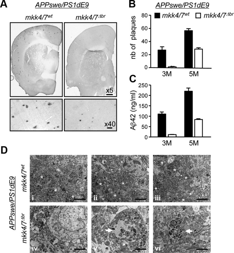
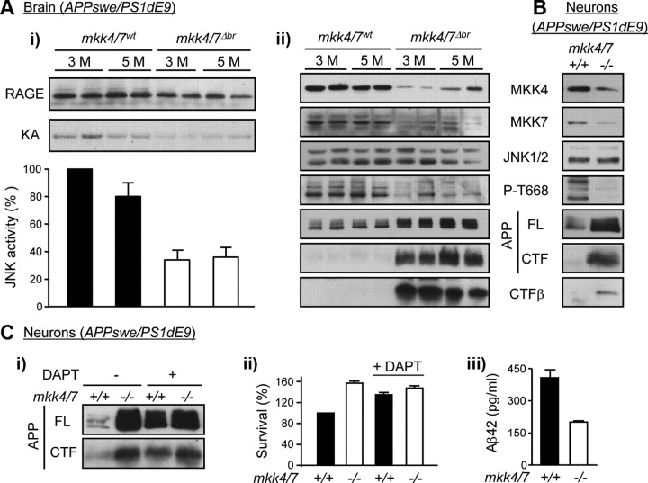
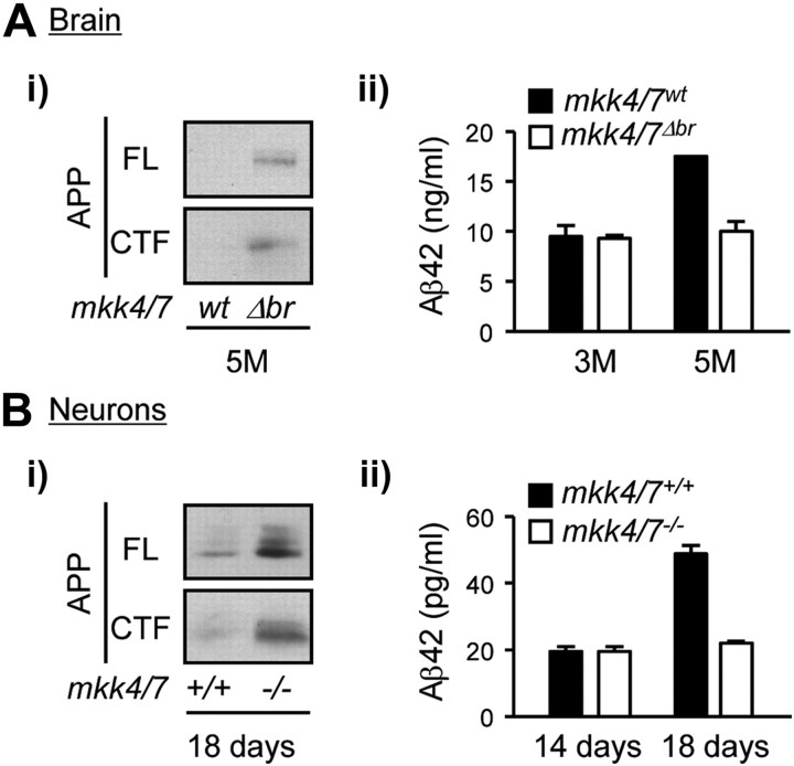
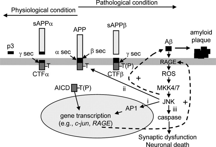
Phosphorylation plays a central role in the dynamic regulation of the processing of the amyloid precursor protein (APP) and the production of amyloid-β (Aβ), one of the clinically most important factors that determine the onset of Alzheimer's disease (AD). This has led to the hypothesis that aberrant Aβ production associated with AD results from regulatory defects in signal transduction. However, conflicting findings have raised a debate over the identity of the signaling pathway that controls APP metabolism. Here, we demonstrate that activation of the c-Jun N-terminal protein kinase (JNK) is essential for mediating the apoptotic response of neurons to Aβ. Furthermore, we discovered that the functional loss of JNK signaling in neurons significantly decreased the number of amyloid plaques present in the brain of mice carrying familial AD-linked mutant genes. This correlated with a reduction in Aβ production. Biochemical analyses indicate that the phosphorylation of APP at threonine 668 by JNK is required for γ-mediated cleavage of the C-terminal fragment of APP produced by β-secretase. Overall, this study provides genetic evidence that JNK signaling is required for the formation of amyloid plaques in vivo. Therefore, inhibition of increased JNK activity associated with aging or with a pathological condition constitutes a potential strategy for the treatment of AD.


Figures

Figure 1.
Figure 1.
The loss of MKK4 and MKK7 protects neurons against Aβ42-induced apoptosis. Cortical neurons displaying homozygous fl mutation for the mkk4 and/or mkk7 alleles and expressing (+) or not (−) CreERT2 were incubated for 72 h with 0.1 μm 4-OHT before being treated with Aβ42 (200 nm) for the indicated times. Where indicated, the cells were pretreated with zVAD (10 μm). A, Protein lysates were analyzed by immunoblot with antibodies against MKK4, MKK7, and JNK. B, E, Immunofluorescence was performed with a specific antibody to MAP2. The immune complex was detected with a secondary antibody conjugated to fluorescein (green). DNA was stained with DAPI (blue). Scale bar, 10 μm. C, Caspase 3 activity was measured by caspase assay. D, Cell survival was measured by MTT assay. The data correspond to the mean ± SE (N = 3).
Figure 2.
Figure 2.
MKK4 and MKK7 are required for Aβ-induced JNK activation. Cortical neurons were incubated with Aβ42 (200 nm) for the indicated times 3 d after 4-OHT treatment to induce gene deletion. Where indicated, the cells were pretreated with NAC (1 mm) or incubated in presence of a neutralizing RAGE antibody (RAGE-AB) (50 μg/ml) before stimulation. A, B, E, Endogenous JNK activity was measured by protein kinase assay. Radioactivity incorporated in the GST-c-Jun substrate was quantified by PhosphorImager. Values are expressed as fold of untreated mkk4/7+/+ sample. The data correspond to the mean ± SE (N = 3) (A, B) or ± SD (N = 2) (E). C, Protein lysates were analyzed by immunoblot with antibodies against MKK4, MKK7, JNK1/2, c-Jun, and active (act) caspase 3. The data are representative of three independent experiments. D, Immunofluorescence was performed with a specific antibody to RAGE. The immune complex was detected with a secondary antibody conjugated to Texas Red (red). DNA was stained with DAPI (blue). Scale bar, 10 μm. F, Caspase 3 activity was measured by caspase assay. The data correspond to the mean ± SE (N = 3).
Figure 3.
Figure 3.
Inactivation of MKK4 and MKK7 in the adult forebrain. One-month-old mkk4/7fl/fl mice carrying (+) or not (−) the CaMKIIcreERT2 transgene were injected with tamoxifen to induce Cre-mediated recombination of the fl alleles. A, One month later, brain sections were analyzed by immunohistochemistry using antibodies against MKK4 or MKK7. Scale bar, 10 μm. HC, Hippocampus; DG, dentate gyrus. B, Protein lysates were analyzed by immunoblot with specific antibodies. The data are representative of two independent experiments. C, Endogenous JNK activity was measured by protein kinase assay. Radioactivity incorporated in the GST-c-Jun substrate was quantified by PhosphorImager. Values are expressed as fold of maximum. The data correspond to the mean ± SD (N = 2 animals).
Figure 4.
Figure 4.
The loss of MKK4 and MKK7 prevents the formation of amyloid plaques. A breeding program was set up to create a mouse model harboring homozygous mkk4fl and mkkk7fl alleles together with a transgene encoding APPswe and PS1dE9, with or without CaMKII-creERT2. Littermates were injected 1 month after birth with tamoxifen, to produce animals lacking MKK4 and MKK7 expression in the brain (mkk4/7Δbr) and suitable controls (mkk4/7wt). A, The brains of 5-month-old mice were analyzed by immunohistochemistry using an antibody to Aβ42. Scale bars: for 5×, 200 μm; 40×, 20 μm. B, The number (nb) of plaques was counted in serial coronal brain sections at 3 and 5 months (M) after birth. C, The amount of Aβ42 was measured by ELISA (Invitrogen). The data correspond to the mean ± SE (N = 3 animals). D, Brain sections were analyzed by EM. The images are from three distinct animals per genotype (i–iii, mkk4/7wt; iv–vi, mkk4/7Δbr). Dystrophic neurites filled with altered mitochondria and residual bodies are clustered around amyloid deposits in mkk4/7wt mice (asterisks), whereas rare isolated dystrophic neurites (arrows) are detected in mkk4/7Δbr animals. Scale bars: i–iv, 2 μm; v, 1 μm; vi, 1.5 μm.
Figure 5.
Figure 5.
MKK4 and MKK7 are required for APP phosphorylation at T668. A, mkk4/7fl/fl littermates carrying the AD disease genes with or without CaMKII-creERT2 were injected 1 month after birth with tamoxifen to produce 3- and 5-month (M)-old animals lacking MKK4 and MKK7 expression in the brain (mkk4/7Δbr) and suitable controls (mkk4/7wt). i, Endogenous JNK activity in brain extracts was measured by protein kinase assay. Radioactivity incorporated in the GST-c-Jun substrate was quantified by PhosphorImager. The autoradiography represents two distinct animals per genotype per time point. The data correspond to the mean ± SE (N = 4 animals). ii, Protein lysates were analyzed by immunoblot with specific antibodies. The data represent two distinct animals per genotype per time point. B, C, Cortical neurons were obtained from mkk4/7fl/fl mice carrying the APPswe/PS1dE9 transgene with (+) or without (−) CaMKII-creERT2 and treated with 4-OHT after 7 DIV to produce mkk4/7/ neurons and suitable controls (mkk4/7+/+). Neurons were used 7 d after the addition of 4-OHT. Where indicated, the cells were treated for 2.5 h with DAPT (1 μm; Sigma-Aldrich) before being harvested. Cell lysates were analyzed by immunoblot using specific antibodies (B, Ci). The data are representative of two experiments. Cell survival was measured by MTT assay (ii). The amount of Aβ42 in cell lysates was measured by ELISA (Invitrogen) (iii). The data correspond to the mean ± SD (N = 2).
Figure 6.
Figure 6.
The amyloidogenic cleavage of endogenous APP requires JNK signaling. A, Brains were obtained from 3- and 5-month (M)-old mkk4/7Δbr mice and control (mkk4/7wt) littermate. B, Cortical neurons lacking MKK4 and MKK7 (mkk4/7/) and suitable controls (mkk4/7+/+) were maintained in culture for 14 or 18 d. Lysates were analyzed by immunoblot using an antibody against APP (i). The amount of Aβ42 was measured by ELISA (IBL International) (ii). The data correspond to the mean ± SD (N = 2).
Figure 7.
Figure 7.
Increased JNK activity induces amyloid plaque formation and neuronal demise. Under normal conditions, Aβ is a relatively minor product in the brain because the nonamyloidogenic processing of APP by α- and γ-secretases prevails. α-secretase-mediated cleavage of APP generates secreted (s) APPα and CTFα. Processing of CTFα by γ secretase produces p3 and an APP intracellular domain (AICD). However, defects in Aβ clearance, associated with aging or a pathological condition, lead to a rise in basal Aβ level. Aβ interacts with RAGE to produce ROS and activates JNK via MKK4 and MKK7. Active JNK phophorylates AP1 transcription factors to regulate gene expression (i), and APP at T668 to enhance the amyloidogenic cleavage of APP (ii). CTFβ generated by β-secretase is processed by γ-secretase to produce a phosphorylated form of AICD, which can translocate to the nucleus (Chang et al., 2006), and more Aβ, which further stimulates JNK activity. Sustained high-level JNK activity in the brain induces caspase activation (iii) causing synaptic dysfunction (D'Amelio et al., 2011) and/or neuronal death. A key therapeutic strategy may be to interfere with the amplification of positive-feedback loops that shifts the balance toward the amyloidogenic pathway leading to increased Aβ42 production and AD pathology. sec, secretase.

References

    1. Akiyama H, Shin RW, Uchida C, Kitamoto T, Uchida T. Pin1 promotes production of Alzheimer's amyloid beta from beta-cleaved amyloid precursor protein. Biochem Biophys Res Commun. 2005;336:521–529. - PubMed
    1. Ando K, Iijima KI, Elliott JI, Kirino Y, Suzuki T. Phosphorylation-dependent regulation of the interaction of amyloid precursor protein with Fe65 affects the production of beta-amyloid. J Biol Chem. 2001;276:40353–40361. - PubMed
    1. Behl C, Davis JB, Lesley R, Schubert D. Hydrogen peroxide mediates amyloid beta protein toxicity. Cell. 1994;77:817–827. - PubMed
    1. Bertram L, Lill CM, Tanzi RE. The genetics of Alzheimer disease: back to the future. Neuron. 2010;68:270–281. - PubMed
    1. Braithwaite SP, Schmid RS, He DN, Sung ML, Cho S, Resnick L, Monaghan MM, Hirst WD, Essrich C, Reinhart PH, Lo DC. Inhibition of c-Jun kinase provides neuroprotection in a model of Alzheimer's disease. Neurobiol Dis. 2010;39:311–317. - PMC - PubMed

Publication types

Substances