Comparative Study
. 2011 Nov 23;31(47):17007-16.
doi: 10.1523/JNEUROSCI.3981-11.2011.

Calcium/calmodulin kinase II in the pedunculopontine tegmental nucleus modulates the initiation and maintenance of wakefulness

Affiliations
Comparative Study

Calcium/calmodulin kinase II in the pedunculopontine tegmental nucleus modulates the initiation and maintenance of wakefulness

Subimal Datta et al. J Neurosci. .

Abstract

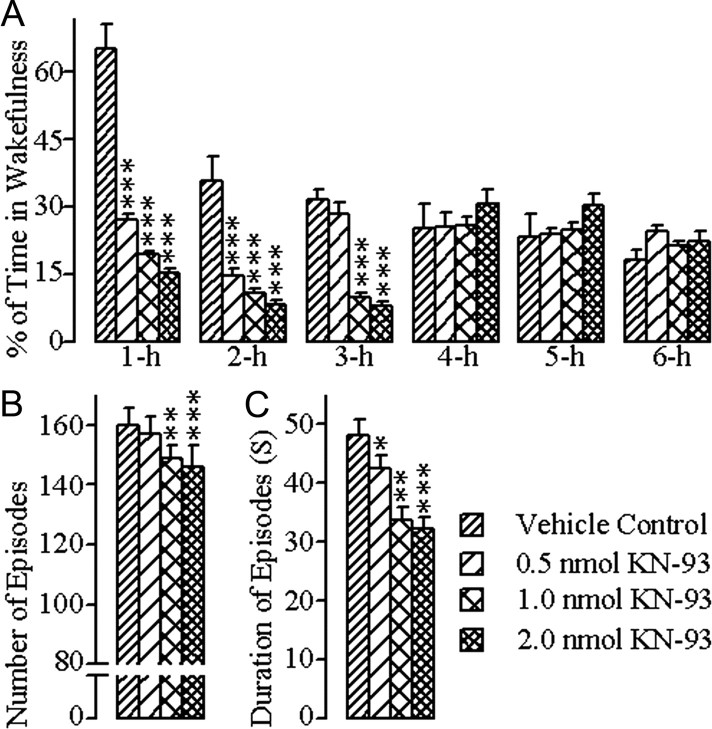
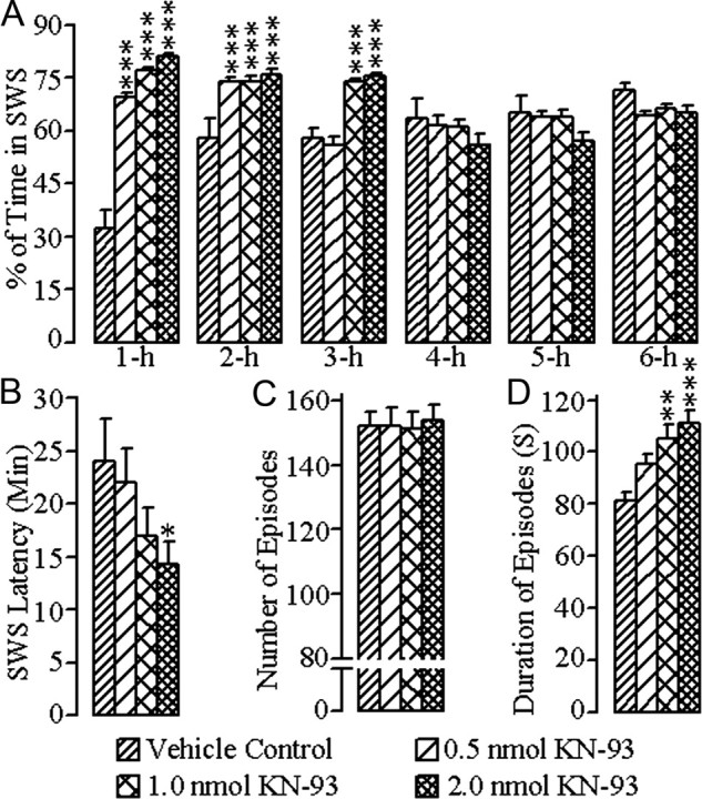
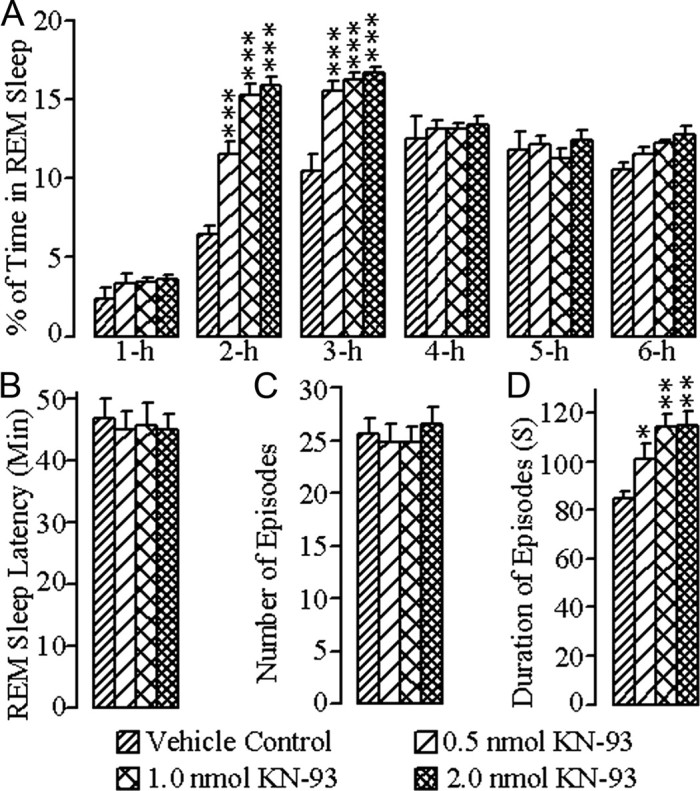
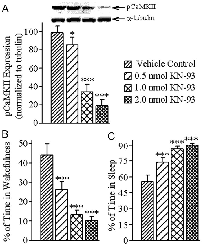
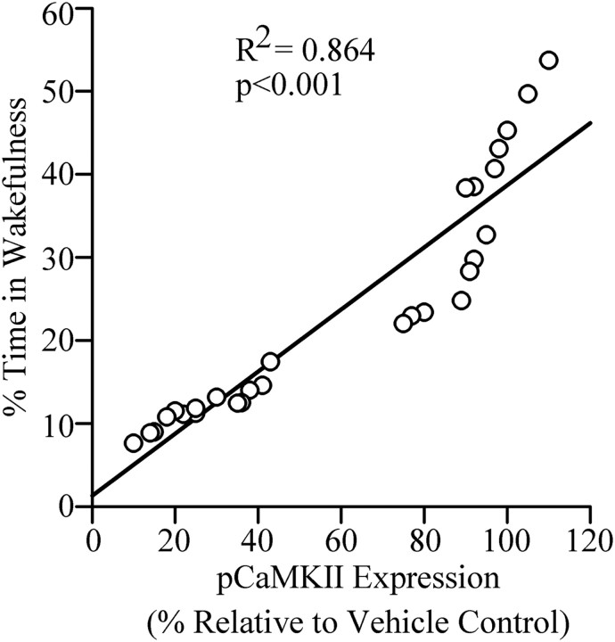
The pedunculopontine tegmentum nucleus (PPT) is critically involved in the regulation of wakefulness (W) and rapid eye movement (REM) sleep, but our understanding of the mechanisms of this regulation remains incomplete. The present study was designed to determine the role of PPT intracellular calcium/calmodulin kinase (CaMKII) signaling in the regulation of W and sleep. To achieve this aim, three different concentrations (0.5, 1.0, and 2.0 nmol) of the CaMKII activation inhibitor, KN-93, were microinjected bilaterally (100 nl/site) into the PPT of freely moving rats, and the effects on W, slow-wave sleep (SWS), REM sleep, and levels of phosphorylated CaMKII (pCaMKII) expression in the PPT were quantified. These effects, which were concentration-dependent and affected wake-sleep variables for 3 h, resulted in decreased W, due to reductions in the number and duration of W episodes; increased SWS and REM sleep, due to increases in episode duration; and decreased levels of pCaMKII expression in the PPT. Regression analyses revealed that PPT levels of pCaMKII were positively related with the total percentage of time spent in W (R(2) = 0.864; n = 28 rats; p < 0.001) and negatively related with the total percentage of time spent in sleep (R(2) = 0.863; p < 0.001). These data provide the first direct evidence that activation of intracellular CaMKII signaling in the PPT promotes W and suppresses sleep. These findings are relevant for designing a drug that could treat excessive sleepiness by promoting alertness.


Figures

Figure 1.
Figure 1.
Effects of vehicle control (saline) and three different concentrations of KN-93 microinjected bilaterally into the PPT on W. These effects were observed during the 6 h wake–sleep recording periods that immediately followed microinjections. A, Bars represent percentages (mean ± SE) of time spent in W. B, Bars represent total number (mean ± SE) of W episodes. C, Bars represent duration (mean ± SE) of W episodes. Asterisks indicate the levels of statistical significance (post hoc tests, Bonferroni post-test) of the differences relative to vehicle control: *p < 0.05; **p < 0.01; ***p < 0.001.
Figure 2.
Figure 2.
Effects of vehicle control (saline) and three different concentrations of KN-93 microinjected bilaterally into the PPT on SWS. These effects were observed during the 6 h wake–sleep recording periods that immediately followed microinjections. A, Bars represent percentages (mean ± SE) of time spent in SWS. B, Bars represent latency (mean ± SE) for the appearance of the first episode of SWS. C, Bars represent total number (mean ± SE) of SWS episodes. D, Bars represent duration (mean ± SE) of SWS episodes. Asterisks indicate the levels of statistical significance (post hoc tests, Bonferroni post-test) of the differences relative to vehicle control: *p < 0.05; **p < 0.01; ***p < 0.001.
Figure 3.
Figure 3.
Effects of vehicle control (saline) and three different concentrations of KN-93 microinjected bilaterally into the PPT on REM sleep. These effects were observed during the 6 h wake–sleep recording periods that immediately followed microinjections. A, Bars represent percentages (mean ± SE) of time spent in REM sleep. B, Bars represent latency (mean ± SE) for the appearance of the first episode of REM sleep. C, Bars represent total number (mean ± SE) of REM sleep episodes. D, Bars represent duration (mean ± SE) of REM sleep episodes. Asterisks indicate the levels of statistical significance (post hoc tests, Bonferroni post-test) of the differences relative to vehicle control: *p < 0.05; **p < 0.01; ***p < 0.001.
Figure 4.
Figure 4.
Effects of vehicle control (saline) and three different concentrations of KN-93 microinjected bilaterally into the PPT on the level of pCaMKII expression in the PPT and percentage of time spent in W and total sleep (TS). A, Representative Western blots of pCaMKII and α-tubulin and data from densitometric analysis of the PPT pCaMKII blots expressed as a percentage of vehicle control in the PPT. All analyses of pCaMKII expression are normalized against α-tubulin. Bars represent levels of PPT pCaMKII expression (mean ± SE). Note that PPT levels of pCaMKII expression decreased in a concentration-dependent manner. B, Bars represent percentages of time (mean ± SE) spent in W during the 3 h period after microinjections. C, Bars represent percentages of time spent in TS (mean ± SE) during the 3 h period after microinjections. Asterisks indicate the levels of statistical significance (post hoc tests, Bonferroni post-test) of the differences relative to vehicle control: *p < 0.05; ***p < 0.001.
Figure 5.
Figure 5.
The relationship between level of pCaMKII expression in the PPT and percentage of time spent in W after bilateral microinjections of vehicle control and three different concentrations of KN-93 into the PPT. Plot of linear regression best fit (solid line) showed a statistically significant positive slope (R2 = 0.864; p < 0.001) between pCaMKII expression and individual animals' total percentage of time spent in W. These data indicate that the percentage of time spent in W after microinjection into the PPT depends positively on the level of pCaMKII expression in the PPT.

References

    1. Bandyopadhya RS, Datta S, Saha S. Activation of pedunculopontine tegmental protein kinase A: a mechanism for rapid eye movement sleep generation in the freely moving rat. J Neurosci. 2006;26:8931–8942. - PMC - PubMed
    1. Capece ML, Lydic R. cAMP and protein kinase A modulate cholinergic rapid eye movement sleep generation. Am J Physiol. 1997;273:R1430–R1440. - PubMed
    1. Chen Y, Luo F, Yang C, Kirkmire CM, Wang ZJ. Acute inhibition of Ca2+/calmodulin-dependent protein kinase II reverses experimental neuropathic pain in mice. J Pharmacol Exp Ther. 2009;330:650–659. - PMC - PubMed
    1. Choi SS, Seo YJ, Shim EJ, Kwon MS, Lee JY, Ham YO, Suh HW. Involvement of phosphorylated Ca2+/calmodulin-dependent protein kinase II and phosphorylated extracellular signal-regulated protein in the mouse formalin pain model. Brain Res. 2006;1108:28–38. - PubMed
    1. Clements JR, Grant S. Glutamate-like immunoreactivity in neurons of the laterodorsal tegmental and pedunculopontine nuclei in the rat. Neurosci Lett. 1990;120:70–73. - PubMed

Publication types

MeSH terms

LinkOut - more resources