Nonsurgical healing of large periradicular lesions using a triple antibiotic paste: A case series
- PMID: 22114375
- PMCID: PMC3220065
- DOI: 10.4103/0976-237X.62519
Nonsurgical healing of large periradicular lesions using a triple antibiotic paste: A case series
Abstract
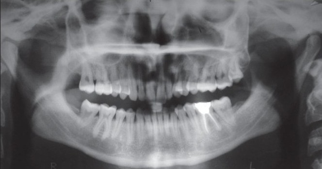
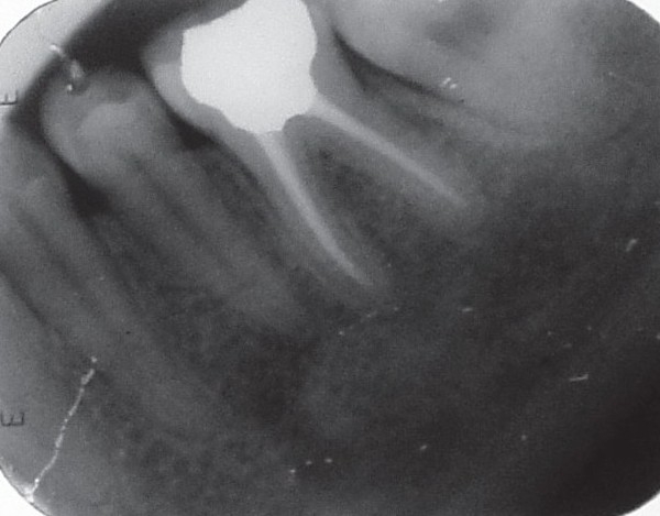
The infection of the root canal system is considered to be a polymicrobial infection, consisting of both aerobic and anaerobic bacteria. Because of the complexity of the root canal infection, it is unlikely that any single antibiotic could result in effective sterilization of the canal. A combination of antibiotic drugs (metronidazole, ciprofloxacin, and minocycline) is used to eliminate target bacteria, which are possible sources of endodontic lesions. Three case reports describe the nonsurgical endodontic treatment of teeth with large periradicular lesions. A triple antibiotic paste was used for 3 months. After 3 months, teeth were asymptomatic and were obturated. The follow-up radiograph of all the three cases showed progressive healing of periradicular lesions. The results of these cases show that when most commonly used medicaments fail in eliminating the symptoms then a triple antibiotic paste can be used clinically in the treatment of teeth with large periradicular lesions.
Keywords: Ciprofloxacin; metronidazole; minocycline; nonsurgical root canal treatment; periradicular lesion; triple antibiotic paste.
Conflict of interest statement
Figures


References
-
- Kakehashi S, Stanley HR, Fitzgerald RJ. The effects of surgical exposure of dental pulps in germ-free and conventional laboratory rats. Oral Surg Oral Med Oral Pathol. 1965:340–9. - PubMed
-
- Sundqvist G. Ecology of the root canal flora. J Endod. 1992;18:427–30. - PubMed
-
- Hoen MM, LaBounty GL, Strittmatter EJ. Conservative treatment of persistent periradicular lesions using aspiration and irrigation. J Endod. 1990;16:182–6. - PubMed
-
- Őztan MD. Endodontic treatment of teeth associated with a large periapical lesion. Int Endod J. 2002;35:73–8. - PubMed
-
- Çalişkan MK. Prognosis of large cyst-like periapical lesions following nonsurgical root canal treatment: A clinical review. Int Endod J. 2004;37:408–16. - PubMed