Ultra-efficient PrP(Sc) amplification highlights potentialities and pitfalls of PMCA technology
- PMID: 22114554
- PMCID: PMC3219717
- DOI: 10.1371/journal.ppat.1002370
Ultra-efficient PrP(Sc) amplification highlights potentialities and pitfalls of PMCA technology
Abstract
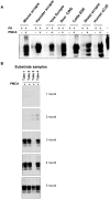
In order to investigate the potential of voles to reproduce in vitro the efficiency of prion replication previously observed in vivo, we seeded protein misfolding cyclic amplification (PMCA) reactions with either rodent-adapted Transmissible Spongiform Encephalopathy (TSE) strains or natural TSE isolates. Vole brain homogenates were shown to be a powerful substrate for both homologous or heterologous PMCA, sustaining the efficient amplification of prions from all the prion sources tested. However, after a few serial automated PMCA (saPMCA) rounds, we also observed the appearance of PK-resistant PrP(Sc) in samples containing exclusively unseeded substrate (negative controls), suggesting the possible spontaneous generation of infectious prions during PMCA reactions. As we could not definitively rule out cross-contamination through a posteriori biochemical and biological analyses of de novo generated prions, we decided to replicate the experiments in a different laboratory. Under rigorous prion-free conditions, we did not observe de novo appearance of PrP(Sc) in unseeded samples of M109M and I109I vole substrates, even after many consecutive rounds of saPMCA and working in different PMCA settings. Furthermore, when positive and negative samples were processed together, the appearance of spurious PrP(Sc) in unseeded negative controls suggested that the most likely explanation for the appearance of de novo PrP(Sc) was the occurrence of cross-contamination during saPMCA. Careful analysis of the PMCA process allowed us to identify critical points which are potentially responsible for contamination events. Appropriate technical improvements made it possible to overcome PMCA pitfalls, allowing PrP(Sc) to be reliably amplified up to extremely low dilutions of infected brain homogenate without any false positive results even after many consecutive rounds. Our findings underline the potential drawback of ultrasensitive in vitro prion replication and warn on cautious interpretation when assessing the spontaneous appearance of prions in vitro.
Conflict of interest statement
The authors have declared that no competing interests exist.
Figures


Similar articles
-
Adaptation of the protein misfolding cyclic amplification (PMCA) technique for the screening of anti-prion compounds.FASEB J. 2024 Jul 31;38(14):e23843. doi: 10.1096/fj.202400614R. FASEB J. 2024. PMID: 39072789 Free PMC article.
-
De novo generation of infectious prions in vitro produces a new disease phenotype.PLoS Pathog. 2009 May;5(5):e1000421. doi: 10.1371/journal.ppat.1000421. Epub 2009 May 15. PLoS Pathog. 2009. PMID: 19436715 Free PMC article.
-
Protein Misfolding Cyclic Amplification Cross-Species Products of Mouse-Adapted Scrapie Strain 139A and Hamster-Adapted Scrapie Strain 263K with Brain and Muscle Tissues of Opposite Animals Generate Infectious Prions.Mol Neurobiol. 2017 Jul;54(5):3771-3782. doi: 10.1007/s12035-016-9945-8. Epub 2016 Jun 4. Mol Neurobiol. 2017. PMID: 27259989
-
Protein misfolding cyclic amplification (PMCA): Current status and future directions.Virus Res. 2015 Sep 2;207:47-61. doi: 10.1016/j.virusres.2014.11.007. Epub 2014 Nov 13. Virus Res. 2015. PMID: 25445341 Review.
-
Prion protein conversion in vitro.J Mol Med (Berl). 2004 Jun;82(6):348-56. doi: 10.1007/s00109-004-0534-3. Epub 2004 Mar 10. J Mol Med (Berl). 2004. PMID: 15014886 Review.
Cited by
-
Correlation between infectivity and disease associated prion protein in the nervous system and selected edible tissues of naturally affected scrapie sheep.PLoS One. 2015 Mar 25;10(3):e0122785. doi: 10.1371/journal.pone.0122785. eCollection 2015. PLoS One. 2015. PMID: 25807559 Free PMC article.
-
Highly infectious prions generated by a single round of microplate-based protein misfolding cyclic amplification.mBio. 2013 Dec 31;5(1):e00829-13. doi: 10.1128/mBio.00829-13. mBio. 2013. PMID: 24381300 Free PMC article.
-
Towards an improved early diagnosis of neurodegenerative diseases: the emerging role of in vitro conversion assays for protein amyloids.Acta Neuropathol Commun. 2020 Jul 25;8(1):117. doi: 10.1186/s40478-020-00990-x. Acta Neuropathol Commun. 2020. PMID: 32711575 Free PMC article. Review.
-
The molecular determinants of a universal prion acceptor.PLoS Pathog. 2024 Sep 10;20(9):e1012538. doi: 10.1371/journal.ppat.1012538. eCollection 2024 Sep. PLoS Pathog. 2024. PMID: 39255320 Free PMC article.
-
Synthesis of high titer infectious prions with cofactor molecules.J Biol Chem. 2014 Jul 18;289(29):19850-4. doi: 10.1074/jbc.R113.511329. Epub 2014 May 23. J Biol Chem. 2014. PMID: 24860097 Free PMC article. Review.
References
-
- Weissmann C. A 'unified theory' of prion propagation. Nature. 1991;352:679–83. - PubMed
-
- Dickinson AG, Outram GW. Genetic aspects of unconventional virus infections: the basis of the virino hypothesis. Ciba Found Symp. 1988;135:63–83. - PubMed
-
- Legname G, Baskakov IV, Nguyen HO, Riesner D, Cohen FE, et al. Synthetic mammalian prions. Science. 2004;305:673–6. - PubMed
Publication types
MeSH terms
Substances
LinkOut - more resources
Full Text Sources
Research Materials

