. 2011 Nov;7(11):e1002391.
doi: 10.1371/journal.ppat.1002391. Epub 2011 Nov 17.

Down-regulation of Shadoo in prion infections traces a pre-clinical event inversely related to PrP(Sc) accumulation

Affiliations

Down-regulation of Shadoo in prion infections traces a pre-clinical event inversely related to PrP(Sc) accumulation

David Westaway et al. PLoS Pathog. 2011 Nov.

Abstract

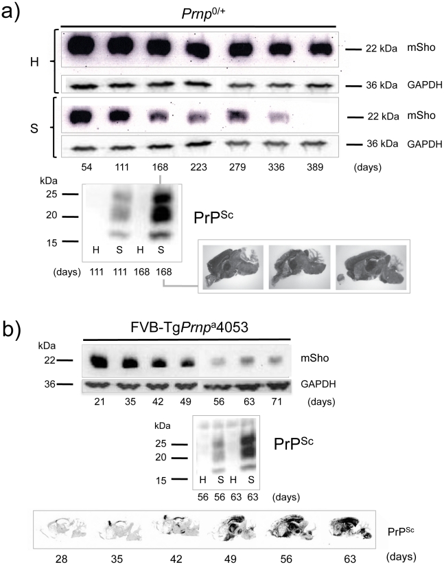
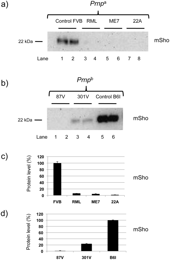
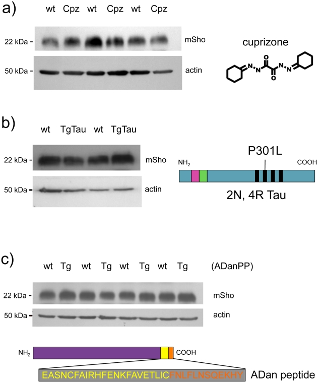
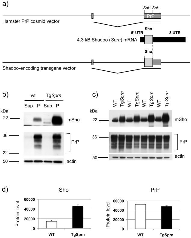
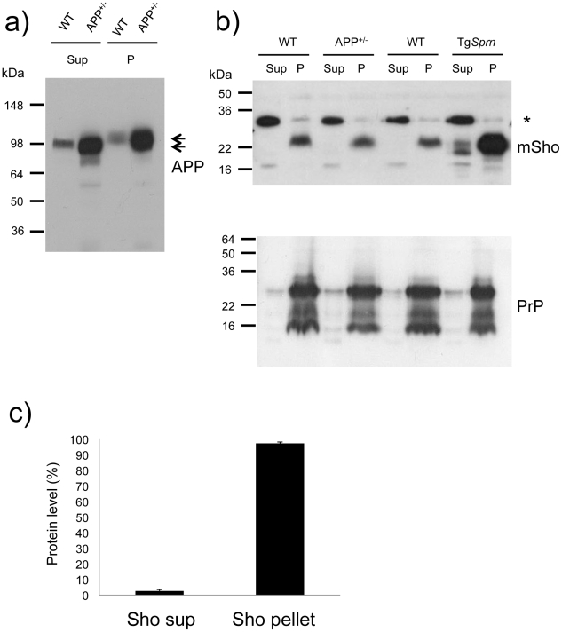
During prion infections of the central nervous system (CNS) the cellular prion protein, PrP(C), is templated to a conformationally distinct form, PrP(Sc). Recent studies have demonstrated that the Sprn gene encodes a GPI-linked glycoprotein Shadoo (Sho), which localizes to a similar membrane environment as PrP(C) and is reduced in the brains of rodents with terminal prion disease. Here, analyses of prion-infected mice revealed that down-regulation of Sho protein was not related to Sprn mRNA abundance at any stage in prion infection. Down-regulation was robust upon propagation of a variety of prion strains in Prnp(a) and Prnp(b) mice, with the exception of the mouse-adapted BSE strain 301 V. In addition, Sho encoded by a TgSprn transgene was down-regulated to the same extent as endogenous Sho. Reduced Sho levels were not seen in a tauopathy, in chemically induced spongiform degeneration or in transgenic mice expressing the extracellular ADan amyloid peptide of familial Danish dementia. Insofar as prion-infected Prnp hemizygous mice exhibited accumulation of PrP(Sc) and down-regulation of Sho hundreds of days prior to onset of neurologic symptoms, Sho depletion can be excluded as an important trigger for clinical disease or as a simple consequence of neuronal damage. These studies instead define a disease-specific effect, and we hypothesize that membrane-associated Sho comprises a bystander substrate for processes degrading PrP(Sc). Thus, while protease-resistant PrP detected by in vitro digestion allows post mortem diagnosis, decreased levels of endogenous Sho may trace an early response to PrP(Sc) accumulation that operates in the CNS in vivo. This cellular response may offer new insights into the homeostatic mechanisms involved in detection and clearance of the misfolded proteins that drive prion disease pathogenesis.


Conflict of interest statement

The authors have declared that no competing interests exist.

Figures

Figure 1
Figure 1. Kinetics of Sho down-regulation in scrapie-infected wt Prnp a/a mouse brain.
a) Time-course of reduction of mature full-length 22 kDa Shadoo protein (mSho) in Prnp a/a mice inoculated with RML scrapie prions and analyzed at the indicated days post-inoculation (06rSH1 antibody). Pair-wise analyses are shown horizontally where “H” designates healthy negative control mice inoculated with control brain whereas “S” designates mice inoculated with the RML isolate of scrapie prions. Glyceraldehyde phosphate dehydrogenase (GAPDH) blot demonstrates similar protein loading for “H” and “S” samples. Western blot analyses show Sho levels in untreated brain samples whereas levels of PrPSc were assessed after PK digestion performed prior to sample electrophoresis and immunoblotting with D18 antibody. Brain histoblot analyses of protease-resistant PrP (D18 antibody) for three mice at the 150-day time-point are shown below the gel analysis, marked “PrPSc”, to demonstrate reproducibility of the protein accumulation. b) Graphical representation of Sho and PrPSc levels in RML-inoculated animals with Sho normalized to 100% for the day 42 time-point in the preclinical phase of disease and with PrPSc normalized to 100% for the day 150 time-point in the clinical phase of disease. In both cases half-maximal change occurred approximately 50 days before the experiment endpoint and at ∼65% elapsed time in disease incubation period.
Figure 2
Figure 2. Kinetics of Sho down-regulation in mice with altered Prnp a gene-dosage.
a) Time-course of reduction of Sho protein in mice hemizygous for Prnp a (Prnp 0/+). Pair-wise analyses (06rSH1 antibody) are presented vertically with the upper row representing control healthy (H) mice and the lower row representing mice inoculated with RML scrapie prions (S), and re-probing as in Figure 1. The time-course was analyzed at different time-points post-inoculation, as shown. The increment in PrPSc levels in scrapie-infected mice (S) detected by western blot analysis is shown for the 111 and 168-day timepoints, with control mice (H) analyzed alongside. Brain histoblot analyses for PrPSc in three mice at the day 168 time-point (pre-clinical phase) are shown as per Figure 1 (D18 antibody). b) Time-course of reduction of Sho protein in TgPrnp a4053 mice overexpressing PrP-A protein. The increment in PrPSc levels between days 56 and 63 is illustrated by western blot analysis. Histoblot time-course (lower panel) demonstrates the accelerated tempo of disease in this transgenic line. PrP analyses were with antibody D18.
Figure 3
Figure 3. Sho levels at disease endpoint in mice infected with different prion strains.
a) Sho protein western blot analysis of brain homogenates from homozygous Prnp a mice infected with different strain isolates and analyzed at clinical phase of prion disease. Biological replicate samples are analyzed for each permutation (lanes 1 and 2, control inoculum; lanes 3 and 4 RML isolate; lanes 5 and 6, ME7 isolate; lanes 7 and 8, 22A isolate). Analyses were with 06rSH1 antibody. “FVB” and “B6I” represent brain samples from animals inoculated with homogenates from healthy FVB and B6I mice, respectively and, thus, comprise negative controls. b) Similar analysis to panel (a) performed with mice of the homozygous Prnp b genotype: (lanes 1 and 2, 87 V isolate; lanes 3 and 4, 301 V isolate; lanes 5 and 6, control inoculum). The position of the mature full-length 22 kDa glycosylated Sho protein (mSho) is shown. c) and d) Densitometric analysis to show percent reduction in Sho in mice inoculated with different prion isolates as per a) and b) versus Sho level in control mice with non-infectious inocula.
Figure 4
Figure 4. Steady-state levels of Sho protein in mice with ‘non-prion’ neurodegenerative syndromes.
Western blot analyses for mature Sho glycoprotein (06rSH1 antibody) are presented for brain homogenates of mice affected by three different disease models. a) Brains samples from cuprizone-treated animals (Cpz) were analyzed after 8 weeks of dosing (0.4% w/w) alongside non-treated controls (wt, wild type). b) Tauopathy. Brain samples from non-Tg (wt) and Tg mice of the TgTau(P301L)23207 line (TgTau) were analyzed at age 545 days. Schematic shows the Tau isoform expressed in this transgenic line. c) Brain samples from 18 month old Tg mice overexpressing the Danish mutant form of BRI2 (known as ADan precursor protein; ADanPP) (Tg) and control non-Tg (wt) mice were analyzed by western blot for Sho protein levels. Prior analyses of tissues from these mice have demonstrated pathologic lesions at this age. A schematic of the mutant BRI2 precursor resulting in extracellular ADan peptide is presented at the bottom of the panel. Wt sequence is represented in yellow while additional sequence present in the mutant is shown in orange. a c) In each case normalized protein loading amounts are demonstrated by re-probing with a β-actin-specific antibody (lower panels).
Figure 5
Figure 5. Sprn and Prnp gene expression time-course in mouse brain.
Mice infected with prions show no significant changes for Prnp and Sprn in three mouse group brains. (a) Prnpa allele-containing mice (FVB, C57Bl/6J) infected with RML or 301 V show almost no changes for Sprn and Prnp, while showing significant changes for GFAP gene transcripts (“Gfap”); (b) Inoculations into Prnpb mice, C57Bl/6.I-1, show similar trends for those genes with profiles in Prnpa allele mice; (c) other mouse strains with various PrPC concentration deriving from different gene copy-numbers infected with RML: FVB-Tg(PrP-A)4053 (Prnp over expressing), Prnp 0/+ hemizygotes and Prnp 0/0control mice that are resistant to prion infections. No significant changes were detected for Sprn and Prnp while significant changes were detected for GFAP only in Prnp 0/+mouse brain. In each case the x-axis shows the incubation time post-inoculation in weeks and the y-axis shows the gene expression change as the log2 median ratio between prion-infected brain samples and control samples.
Figure 6
Figure 6. TgSprn transgenic mice and transgene-encoded Sho protein.
a) Use of the cos.Tet transgene vector to drive Sho protein expression under the control of the Syrian hamster PrP transcriptional unit. A cDNA fragment encoding the wt mouse Sho ORF and devoid of native Sprn 5′ UTR and 3′ UTR mRNA sequences was used for this purpose. b) Upper panel: Blot analysis of supernatant (Sup) and pellet fractions (P) from a 100,000xg centrifugation of brain homogenates of wt and TgSprn24551 mice (N-terminal antibody 06rSH1). Homogenates were prepared in isotonic sucrose. Lower panel: Re-analysis of the same samples with PrP-specific Sha31 antibody indicated a similar pattern of fractionation. c) Upper panel: Comparison of Sho protein (06rSH1 antibody) levels in a 100,000xg pellet in a litter of TgSprn24551 (TgSprn) mice and their non-Tg (wt) littermates. Lower panel: Re-analysis of the same samples with Sha 31 PrP-specific antibody indicates no distinction in the levels of sedimented PrP from Tg and non-Tg animals. Age of analysis of all mice was 95 days. d) Densitometric analysis of the data presented in panel (c) for Sho and PrP, respectively. Protein level on y-axis is in arbitrary units. The degree of overexpression seen in TgSprn24551 mice was estimated at 2.5 x.
Figure 7
Figure 7. Analysis of DEA extracted APP and Sho from mouse brain.
a) Diethylamine (DEA) extraction for wild type (wt) and TgAPP+/- (APP+/-) mouse brains successfully separates the non-integral forms of APP, secreted APP, “sAPP”, as seen in the supernatants (Sup), from the membrane-associated holoprotein that remains in the pellet fraction (P). The TgAPP transgenic line analyzed here corresponds to TgCRND8 mice with ∼ 5x overexpression of APP695 holoprotein. Arrows distinguish the molecular weight difference between processed (extractable) and full-length APP, with electrophoretic mobility of ∼100 and 120 kDa, respectively. b) Upper panel: Unlike the situation for APP, DEA extraction for wild type (wt) and TgAPP+/- (APP+/-) mouse brains fails to release detectable levels of Sho into the supernatant fraction (Sup), and signal remained predominantly in the membrane-associated pellet (P). Lower panel: this demonstrates normalized sample loading by probing for PrP (Sha 31 antibody), with these replica samples being PNGaseF digested to display (from top to bottom) full-length as well as C2 and C1 proteolytic fragments. The asterisk indicates a non-specific band detected with the polyclonal Sho antiserum 06rSH1. c) Densitometric quantitation of data in (b), upper panel, indicating 97 ±1% SEM of the Sho protein in brain samples of wt mice is present in the pellet fraction. Ages of the presented TgAPP and TgSprn24551 mice were 8 and 9 months, respectively.
Figure 8
Figure 8. Prion infection assessed in TgSprn mice.
a) Survival curves of transgenic (TgSprn) and non-transgenic (Non-Tg) genotypes inoculated with RML prions are presented. Using a log-rank (Mantel Cox) test, the chi-square value was 0.24 and the P value 0.6 (not significant). b) Histopathological analysis of infected animals (hematoxylin and eosin stain of hippocampal neurons and the corpus callosum, intracerebral route of inoculation). Transgenic (TgSprn) and non-transgenic (Non-Tg) genotypes are shown. c). Western blot analysis of brain homogenates from groups of four control non-transgenic mice (upper) and four TgSprn mice (lower) with and without prion infection (RML prion isolate). Animals were in the clinical phase of disease and analysis was performed with the anti-Sho N-terminal antibody, 06rSH1. Bar graphs represent percent remaining in each case, normalized to the uninfected control animals as 100%. The percentage diminution in mSho in both cases was over 90% and with the extent of Sho down-regulation not being significantly different between non-Tg and Tg animals. “inf” signifies infected. A cross-reactive band is indicated with an asterisk and also illustrates similar sample loadings between infected and uninfected animals. d) Blot analysis of protease-resistant PrP from non-Tg and TgSprn24551 mice (anti-PrP antibody D18).
Figure 9
Figure 9. Sho protein in chronically infected SMB cells.
a) Prion-infected SMB cells (SMB Sc) or pentosan sulfate cured SMB cells (SMB-PS) were acutely transfected with a control cDNA expression vector (empty vector, e.v.) or the same plasmid vector bearing a cDNA insert encoding wt mouse Sho (Sho). Upper panel: Cell lysates were prepared from cells 24 hours after transfection and were immunoblotted with a C-terminal anti-Sho antibody 06SH3a. Lower panel: A similar transfection efficiency into healthy and infected cells was established by probing for expression of neomycin phosphotransferase encoded within both varieties of plasmid vector (“NPT II”). C1 is a C-terminal fragment of Sho. b) The presence of PrPSc in infected SMB cultures was verified by protease digestion. Two PK resistant fragments co-migrated with fragments present in undigested lysates of infected cells, but not those of uninfected cells (open arrowheads; see also Figure S1). c) Conditioned medium was harvested from the transfected cells, acetone precipitated and then western blotted for the presence of the Sho glycoprotein with 06SH3a antibody.

Similar articles

Cited by

References

    1. Bruce ME, Will RG, Ironside JW, McConnell I, Drummond D, et al. Transmissions to mice indicate that "new variant" CJD is caused by the BSE agent. Nature. 1997;389:498–501. - PubMed
    1. Manson JC, Cancellotti E, Hart P, Bishop MT, Barron RM. The transmissible spongiform encephalopathies: emerging and declining epidemics. Biochem Soc Trans. 2006;34:1155–1158. - PubMed
    1. Westaway D, Goodman PA, Mirenda CA, McKinley MP, Carlson GA, et al. Distinct prion proteins in short and long scrapie incubation period mice. Cell. 1987;51:651–662. - PubMed
    1. Hunter N, Dann JC, Bennett AD, Somerville RA, McConnell I, et al. Are Sinc and the PrP gene congruent? Evidence from PrP gene analysis in Sinc congenic mice. J Gen Virol. 1992;73:2751–2755. - PubMed
    1. Moore RC, Hope J, McBride PA, McConnell I, Selfridge J, et al. Mice with gene targetted prion protein alterations show that Prnp, Sinc and Prni are congruent. Nat Genet. 1998;18:118–125. - PubMed

Publication types