. 2011;6(11):e27520.
doi: 10.1371/journal.pone.0027520. Epub 2011 Nov 17.

Viral single-strand DNA induces p53-dependent apoptosis in human embryonic stem cells

Affiliations

Viral single-strand DNA induces p53-dependent apoptosis in human embryonic stem cells

Matthew L Hirsch et al. PLoS One. 2011.

Abstract

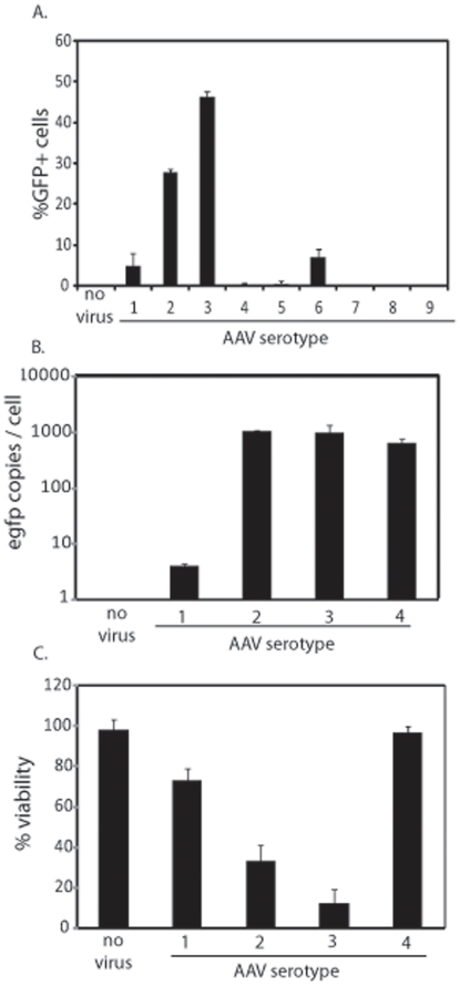
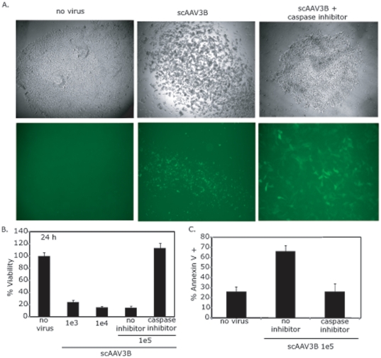
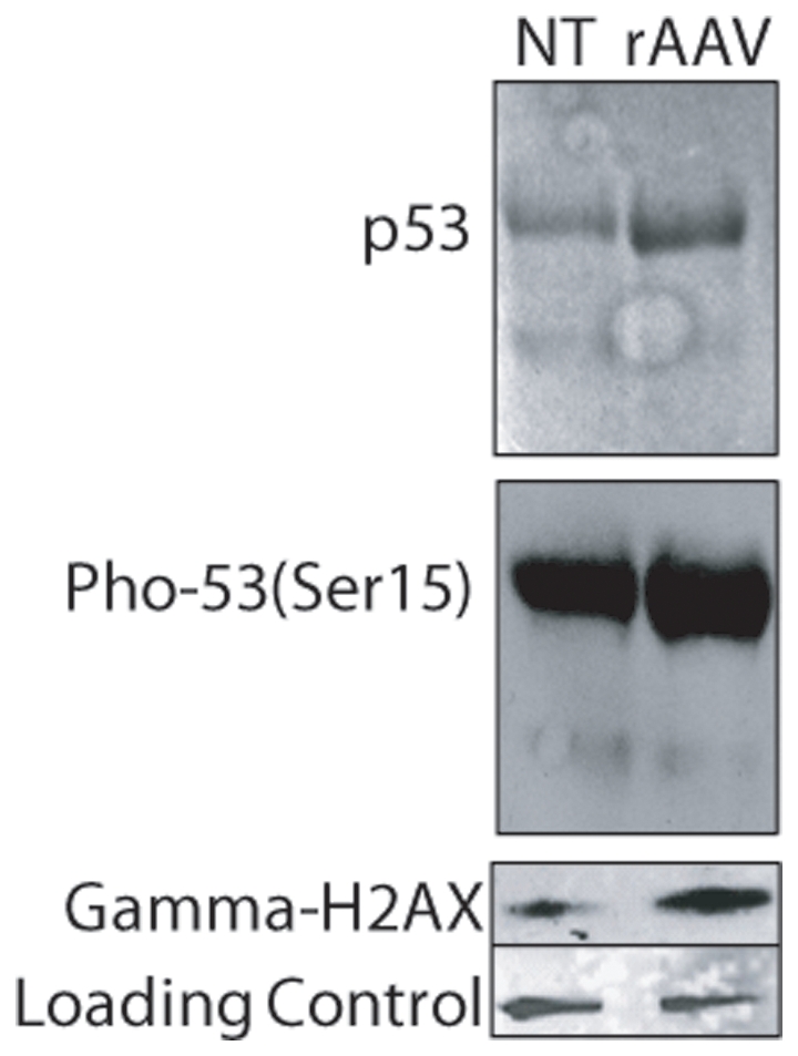
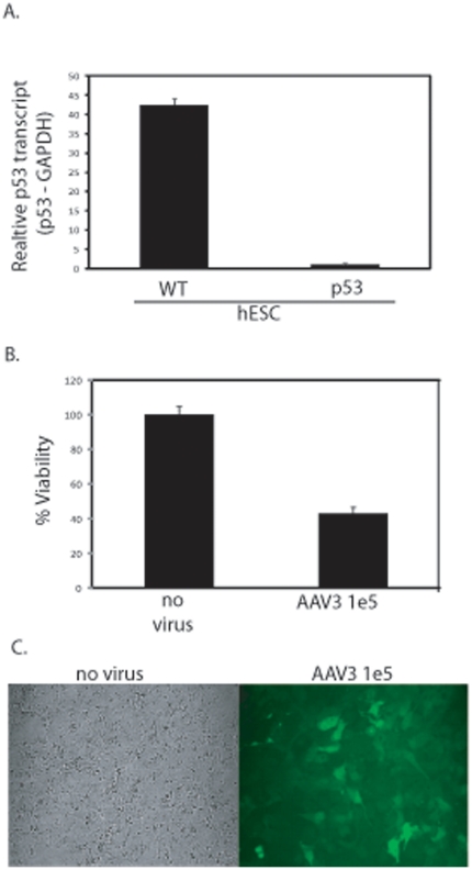
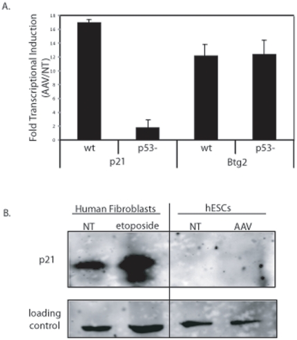
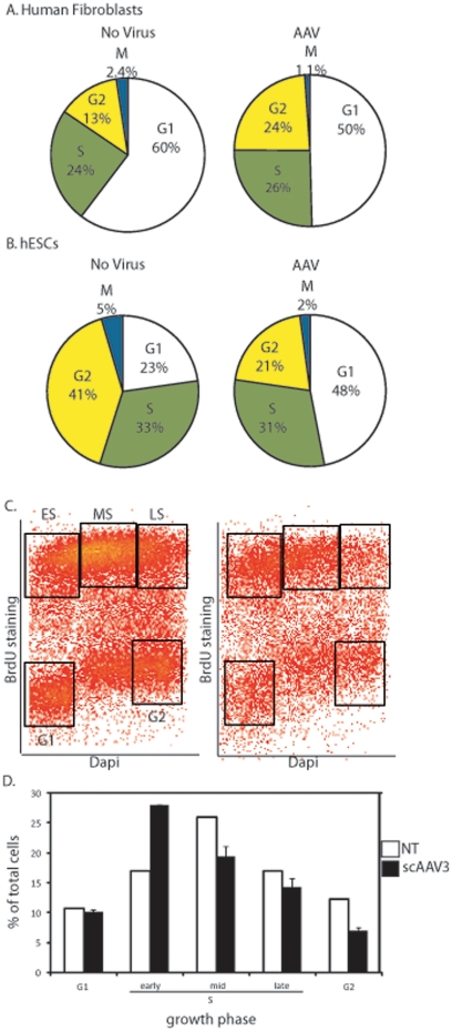
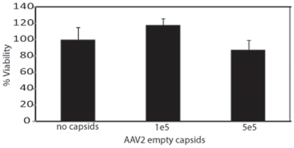
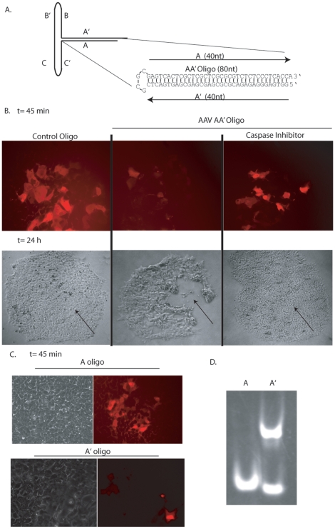
Human embryonic stem cells (hESCs) are primed for rapid apoptosis following mild forms of genotoxic stress. A natural form of such cellular stress occurs in response to recombinant adeno-associated virus (rAAV) single-strand DNA genomes, which exploit the host DNA damage response for replication and genome persistence. Herein, we discovered a unique DNA damage response induced by rAAV transduction specific to pluripotent hESCs. Within hours following rAAV transduction, host DNA damage signaling was elicited as measured by increased gamma-H2AX, ser15-p53 phosphorylation, and subsequent p53-dependent transcriptional activation. Nucleotide incorporation assays demonstrated that rAAV transduced cells accumulated in early S-phase followed by the induction of apoptosis. This lethal signaling sequalae required p53 in a manner independent of transcriptional induction of Puma, Bax and Bcl-2 and was not evident in cells differentiated towards a neural lineage. Consistent with a lethal DNA damage response induced upon rAAV transduction of hESCs, empty AAV protein capsids demonstrated no toxicity. In contrast, DNA microinjections demonstrated that the minimal AAV origin of replication and, in particular, a 40 nucleotide G-rich tetrad repeat sequence, was sufficient for hESC apoptosis. Our data support a model in which rAAV transduction of hESCs induces a p53-dependent lethal response that is elicited by a telomeric sequence within the AAV origin of replication.


Conflict of interest statement

Competing Interests: The authors have declared that no competing interests exist.

Figures

Figure 1
Figure 1. Recombinant AAV Transduction of hESCs.
The indicated AAV serotypes were packaged with a self-complementary CMV-egfp genome and used to infect human embryonic stem cells (hESCs) at 100,000 viral genomes per cell. 24 h post-infection (post-infection) cells were harvested and GFP+ cells were quantitated by flow cytometry (A.). Treated cells were also analyzed for intracellular transgene copy number normalized to the lamin B gene (B). Cell viability was also measured under the indicated conditions via dye exclusion (C) and significant decreases (p-value<0.05) were noted in all cases (compared to no virus) except with AAV4 treatment (p-value = 0.44). The results are averaged from at least 6 replicates for each treatment group and the standard deviation is depicted.
Figure 2
Figure 2. Recombinant AAV Transduction of hESCs induces Apoptosis.
Human embryonic stem cells (hESCs) were treated with 100,000 AAV3B particles packaged with a self-complementary CMV-egfp genome. 24 h post-infection (p.i.) cellular morphology and GFP fluorescence was observed by microscopy in the presence or absence of a pan-caspase inhibitor (A). Cell viability of hESCs treated with the indicated amount of the scAAV3B-egfp vector described above was analyzed by dye exclusion 24 h post-infection (B). Significant decreases (p-value<0.05) were noted in all cases except when the caspase inhibitor was used (p-value>0.1). hESCs treated as described were analyzed for annexin V staining 8 h post-infection (C). A significant increase (p-value<0.05) was noted for cells treated with only rAAV3B. The data in (B) and (C) represents the average from at least 6 replicates per treatment group and the standard deviation is depicted as well.
Figure 3
Figure 3. Recombinant AAV Transduction Activates a DNA Damage Response in hESCs.
Total protein from scAAV3b-egfp transduced hESCs was harvested 8 h post-infection. Western blotting analysis was performed using the indicated antibodies.
Figure 4
Figure 4. Recombinant AAV-Induced hESC Apoptosis is p53-Dependent.
A polyclonal p53 deficient H9 hES cell line was constructed using lenti-viral transduction of a p53-specific shRNA cassette. After selection, total RNA was harvested, converted to cDNA and the relative abundance of p53 transcript, compared to the GAPDH housekeeping transcript, was determined by Q-PCR (A). The parental H9 hESCs were used as a wild type control (A). scAAV3B-egfp transduction of p53 deficient hESCs at a MOI of 100,000 was performed. 24 h post-infection viability was determined by dye exclusion (B) microscopy (C). The results in (A) and (B) are averaged from at least 6 replicates for each treatment group and the standard deviation is depicted. The differences noted in both (A) and (B) have p-values<0.005).
Figure 5
Figure 5. p53 Transactivation Occurs Following Recombinant AAV Infection.
(A) Fold induction of p21 and Btg2 transcripts via quantitative PCR following rAAV infection. Data is presented as the value determined for hESCs infected with 100,000 AAV3B vectors 8 h post-infection divided by the transcript amount for hESCs not treated. Values were determined in both H9s and H9s with p53 knocked down as described in text (p53-). A significant difference was noted only for p21 transcript levels in the absence of p53 (p-value<0.005). B) Western blot analysis of p21 abundance following rAAV infection (8 h post-infection). Human fibroblasts treated with the DNA damaging agent etoposide was used as an antibody control. The results are averaged from at least 3 replicates for each treatment group and the standard deviation is depicted.
Figure 6
Figure 6. AAV Infection of hESCs Induces Early S-phase Accumulation.
A) Normal human fibroblasts treated with 100,000 AAV3B vectors were treated 6 h post-infection with nocodazole. 2 h later cells were harvested and stained with DAPI and a mitotic marker as described in the text. Flow cytometry allowed the determination of cells in the indicated growth phases; however by this assay early S-phase and G1 cells are not distinguished. B) The method described above was performed on hESCs and processed in the same manner. C) hESCs treated with AAV as described above were given EdU 6 h post-infection. 2 h later cells were harvest, stained with DAPI and analyzed using flow cytometry. A representative dot plot is shown with the boxes drawn to represent different growth phases. D) Average quantitation of cells depicted in C). Collectively, the results are averages from at least 3 independent experiments and the standard deviation is depicted (* indicates p-value<0.005).
Figure 7
Figure 7. AAV empty capsid transduction of hESCs is non-toxic.
AAV2 capsids were administered to hESCs at the indicated dose. Cell viability was determined after 24 h by dye exclusion. The average of at least 6 replicates is presented with the standard deviation. No significant difference (p-value>0.1) was noted when treatment groups were compared to the no capsid control).
Figure 8
Figure 8. AAV Inverted Terminal Repeats Induce Apoptosis in hESCs.
A) Cartoon depiction of the AAV inverted terminal repeat. The experimental oligonucleotides are also depicted as AA' (80-mer), A (39-mer) and A' (39-mer). B) hESCs were injected with AA' or a control oligonucleotide with no AAV sequence under the indicated conditions. The DNA solution contained rhodamine allowing visualization of injected cells (red). Microscopy was performed at the indicated time points. C) DNA microinjections of the A or A' oligonucleotides as described above. D) Native gel analysis of the A and A' oligonucleotides as described in the text.

References

    1. Dravid G, Ye Z, Hammond H, Chen G, Pyle A, et al. Defining the role of Wnt/beta-catenin signaling in the survival, proliferation, and self-renewal of human embryonic stem cells. Stem Cells. 2005;10:1489–501. - PubMed
    1. Qin H, Yu T, Qing T, Liu Y, Zhao Y, et al. Regulation of apoptosis and differentiation by p53 in human embryonic stem cells. J Biol Chem. 2007;8:5842–52. - PubMed
    1. Grandela C, Pera MF, Wolvetang EJ. p53 is required for etoposide-induced apoptosis of human embryonic stem cells. Stem Cell Res. 2007;2:116–28. - PubMed
    1. Wang Y, Blelloch R. Cell cycle regulation by MicroRNAs in embryonic stem cells. Cancer Res. 2009;10:4093–6. - PMC - PubMed
    1. Raj K, Ogston P, Beard P. Virus-mediated killing of cells that lack p53 activity. Nature. 2001;412:914–7. - PubMed

Publication types

MeSH terms