Review
. 2012 Mar 15;16(6):506-23.
doi: 10.1089/ars.2011.4260.

Protein glutathionylation in the regulation of peroxiredoxins: a family of thiol-specific peroxidases that function as antioxidants, molecular chaperones, and signal modulators

Affiliations
Review

Protein glutathionylation in the regulation of peroxiredoxins: a family of thiol-specific peroxidases that function as antioxidants, molecular chaperones, and signal modulators

Ho Zoon Chae et al. Antioxid Redox Signal. .

Abstract

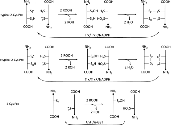
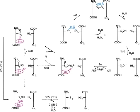
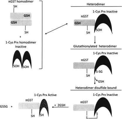
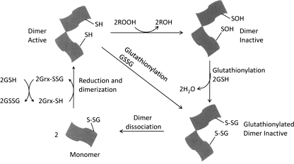
Significance: Reversible protein glutathionylation plays an important role in cellular regulation, signaling transduction, and antioxidant defense. This redox-sensitive mechanism is involved in regulating the functions of peroxiredoxins (Prxs), a family of ubiquitously expressed thiol-specific peroxidase enzymes. Glutathionylation of certain Prxs at their active-site cysteines not only provides reducing equivalents to support their peroxidase activity but also protects Prxs from irreversible hyperoxidation. Typical 2-Cys Prx also functions as a molecular chaperone when it exists as a decamer and/or higher molecular weight complexes. The hyperoxidized sulfinic derivative of 2-Cys Prx is reactivated by sulfiredoxin (Srx). In this review, the roles of glutathionylation in the regulation of Prxs are discussed with respect to their molecular structure and functions as antioxidants, molecular chaperones, and signal modulators.

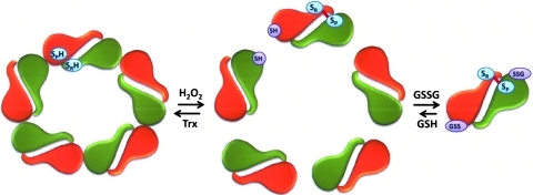
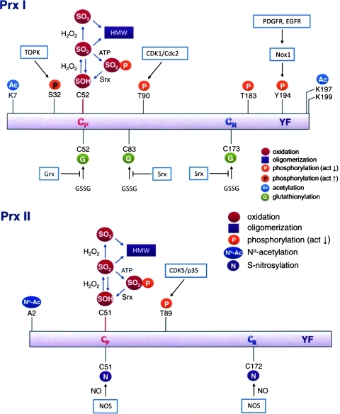
Recent advances: Recent findings reveal that glutathionylation regulates the quaternary structure of Prx. Glutathionylation of Prx I at Cys(83) converts the decameric Prx to its dimers with the loss of chaperone activity. The findings that dimer/oligomer structure specific Prx I binding proteins, e.g., phosphatase and tensin homolog (PTEN) and mammalian Ste20-like kinase-1 (MST1), regulate cell cycle and apoptosis, respectively, suggest a possible link between glutathionylation and those signaling pathways.

Critical issues: Knowing how glutathionylation affects the interaction between Prx I and its nearly 20 known interacting proteins, e.g., PTEN and MST1 kinase, would reveal new insights on the physiological functions of Prx.

Future directions: In vitro studies reveal that Prx oligomerization is linked to its functional changes. However, in vivo dynamics, including the effect by glutathionylation, and its physiological significance remain to be investigated.


Figures

FIG. 1.
FIG. 1.
Peroxidase catalytic mechanisms of peroxiredoxin (Prx). All of the subfamily members of Prx are obligatory dimers. Typical 2-Cys subfamily members form two intermolecular disulfide bonds by resolving the sulfenic derivative (Cys-SOH) of peroxidatic Cys. Atypical 2-Cys Prx forms one intramolecular disulfide bond per each monomer, despite the fact that it exists in an antiparalleled dimer like typical 2-Cys Prx. Whereas, 1-Cys Prx does not form a disulfide due to an unavailability of proximal Cys residue for resolving. Glutathione with π isoform of glutathione S-transferase (π-GST) is required for the resolution of peroxidatic Cys-OH.
FIG. 2.
FIG. 2.
Quaternary structure of Prxs. (A) Dimeric structure of rat isoform of human Prx I with the disulfide bond between SP and SR (ball and stick with sulfur atom colored yellow). This figure is based on a figure from Hirotsu et al. (47). (B) Decameric structure of human Prx I adopted from a figure from Schroder et al. (115). (To see this illustration in color the reader is referred to the web version of this article at www.liebertonline.com/ars).
FIG. 3.
FIG. 3.
Schematic representation of the active site of H2O2-bound Prx. Hydrogen bonding, involving both the protein backbone and side-chain atoms of the conserved four amino acid residues (Pro, Thr, Arg, and Glu/Gln/His) stabilizes the thiolate anion of the peroxidatic cysteine (Cp) and activates the H2O2 molecule for the nucleophilic attack by the thiolate anion. For the Prx-bound H2O2 molecule, the oxygen atoms OA and OB represent the electrophilic center and the atom to be displaced, respectively. This figure is based on a figure from Hall et al. (45).
FIG. 4.
FIG. 4.
Ribbon diagram of a monomer of human Prx V. This structure shows a possible conformation forming an intramolecular disulfide bond between the CP (Cys47) and Cys151. This figure is based on a figure from Evrard et al. (31). (To see this illustration in color the reader is referred to the web version of this article at www.liebertonline.com/ars).
FIG. 5.
FIG. 5.
Peroxidase catalysis, hyperoxidation, and retroreduction mechanism of eukaryotic typical 2-Cys peroxiredoxin. Disulfide bond between peroxidatic and resolving Cys residues (Cys-SP-SR-Cys) is reduced to sulfhydryls by the use of thioredoxin (Trx). As a consequence, the distance between SP and SR extends to 14 Å. The sulfenic intermediate (Cys-SP-OH) is formed by reducing H2O2 to water. When the H2O2 level is low (usually <10 μM), Cys-SP-OH persists until approaching Cys-SRH within the disulfide bonding radius, which requires local unfolding of the structure. When the H2O2 level increases, Cys-SP-OH reacts with an additional H2O2 molecule to form a sulfinic intermediate (Cys-SPO2), consequently Prx loses its peroxidase activity until retroreduced to Cys-SPH. Retroreduction of Cys-SPO2 starts with the formation of sulfinic phosphoryl ester (Cys-(SP=O)-OPO32−), which is catalyzed by sulfiredoxin (Srx) in the presence of ATP and Mg2+. The Srx-Prx thiosulfinate (Prx-Cys-(SP=O)-S-Cys-Srx) intermediate is formed by releasing inorganic phosphate. Reduction of this thiosulfinate with glutathione (GSH) or Trx restores the hyperoxidized Prx to normal peroxidase catalytic intermediate (Cys-SP-OH). Several reducing pathways from Cys-SP-OH to Cys-SPH have been proposed involving GSH and/or Trx in the presence of Srx; however, the preference or detailed mechanism of the reaction remains to be elucidated. The dashed arrows indicate unconfirmed but possible reaction path through which GSH directly attacks the sulfinylphosphate intermediate to form Prx-(Sp=O)-SG and SrxS intermediates. The scheme shows the mechanistic pathway of one of two active sites in the dimeric structure of typical 2-Cys Prx. (To see this illustration in color the reader is referred to the web version of this article at www.liebertonline.com/ars).
FIG. 6.
FIG. 6.
Protein glutathionylation of Prx I induces a decamer to dimer switch. Glutathionylation induces a structural change in Prx I and leads to the dissociation of its decamer to dimers. (To see this illustration in color the reader is referred to the web version of this article at www.liebertonline.com/ars).
FIG. 7.
FIG. 7.
Post-translational modifications of human Prx I and Prx II. Sulfhydryls of peroxidatic cysteine residue (CP) of both Prx I and Prx II can be further oxidized to reversible sulfinic acid or irreversible sulfonate acid derivatives. Both forms of the hyperoxidized Prxs shift their quaternary structure equilibrium in favor of the higher-molecular weight (HMW) structures. Three Cys residues, CP, CR, and C83, in Prx I are the sites of glutathionylation, which is negatively regulated by glutaredoxin or Srx. Both CP and CR of Prx II can be S-nitrosylated by NO generated by NO synthase (NOS). Nα acetylation is found on the penultimate Ala residue of Prx II, and N-acetylation of Lys residues in Prx I at K7, K197, and K199 have been identified. Phosphorylation at T90, T183, S32, and Y194 in Prx I and at T89 in Prx II has been observed. The effects of these phosphorylations on the peroxidase activity are indicated. CDK/Cdc; EGFR, epidermal growth factor receptor; Grx, glutaredoxin; Nox, NADPH oxidase; PDGFR, platelet-derived growth factor receptor; TOPK, T-cell–originated protein kinase. (To see this illustration in color the reader is referred to the web version of this article at www.liebertonline.com/ars).
FIG. 8.
FIG. 8.
A mechanistic scheme depicting the reduction of ROOH catalyzed by PGdx is mediated by the GSH redox cycle. PGdx is a chimeric enzyme from Haemophilus influenza Rd which consists of the N-terminal peroxiredoxin (light section) and the C-terminal glutaredoxin (gray section) region. The reaction steps 1 to 5 were proposed by Pauwels et al. (92) to explain their observed data, while the steps represented by the dotted lines were proposed by Rouhier et al. (109) for the Grx-reducible poplar Prx and adopted here.
FIG. 9.
FIG. 9.
A mechanistic scheme depicting activation of the oxidized 1-Cys Prx by πGST and GSH. The activation involves the formation of a heterodimer between the GSH bound πGST and the oxidized 1-Cys Prx, which leads to the formation of a glutathionylated 1-Cys Prx intermediate and follows with the formation of a disulfide bonded heterodimer prior to its reduction by GSH.
FIG. 10.
FIG. 10.
Proposed peroxidase mechanism for the 1-Cys D Prx. ROOH oxidizes the SP of 1-Cys D Prx to form an inactive sulfenic dimer. Glutathionylation of this inactive dimer induces a conformational change and causes it to dissociate into monomers. The glutathionylated monomers are reduced by the glutaredoxin/glutathione system to complete the catalytic cycle.
FIG. 11.
FIG. 11.
Model of peroxiredoxin VI structure. It shows the peroxidatic Cys 47 (purple); the catalytic center for the peroxidase activity; and Ser 32 (red), His 26 (yellow), and Asp 140 (blue), the catalytic triad of the phospholipase A2 active site. The figure is based on a figure reported by Manevich et al. (76). (To see this illustration in color the reader is referred to the web version of this article at www.liebertonline.com/ars).

References

    1. Adachi T. Pimentel DR. Heibeck T. Hou X. Lee YJ. Jiang B. Ido Y. Cohen RA. S-glutathiolation of Ras mediates redox-sensitive signaling by angiotensin II in vascular smooth muscle cells. J Biol Chem. 2004;279:29857–29862. - PubMed
    1. Alger HM. Williams DL. The disulfide redox system of Schistosoma mansoni and the importance of a multifunctional enzyme, thioredoxin glutathione reductase. Mol Biochem Parasitol. 2002;121:129–139. - PubMed
    1. Aslund F. Ehn B. Miranda-Vizuete A. Pueyo C. Holmgren A. Two additional glutaredoxins exist in Escherichia coli: glutaredoxin 3 is a hydrogen donor for ribonucleotide reductase in a thioredoxin/glutaredoxin 1 double mutant. Proc Natl Acad Sci U S A. 1994;91:9813–9817. - PMC - PubMed
    1. Bae YS. Kang SW. Seo MS. Baines IC. Tekle E. Chock PB. Rhee SG. Epidermal growth factor (EGF)-induced generation of hydrogen peroxide. Role in EGF receptor-mediated tyrosine phosphorylation. J Biol Chem. 1997;272:217–221. - PubMed
    1. Barrett WC. DeGnore JP. Keng YF. Zhang ZY. Yim MB. Chock PB. Roles of superoxide radical anion in signal transduction mediated by reversible regulation of protein-tyrosine phosphatase 1B. J Biol Chem. 1999;274:34543–34546. - PubMed

Publication types