Review
. 2012 Dec 1;17(11):1610-55.
doi: 10.1089/ars.2011.4109. Epub 2012 Jan 18.

Redox proteomics in selected neurodegenerative disorders: from its infancy to future applications

Affiliations
Review

Redox proteomics in selected neurodegenerative disorders: from its infancy to future applications

D Allan Butterfield et al. Antioxid Redox Signal. .

Abstract

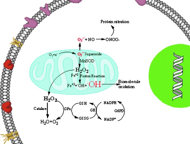
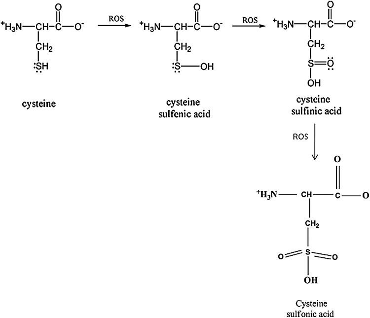
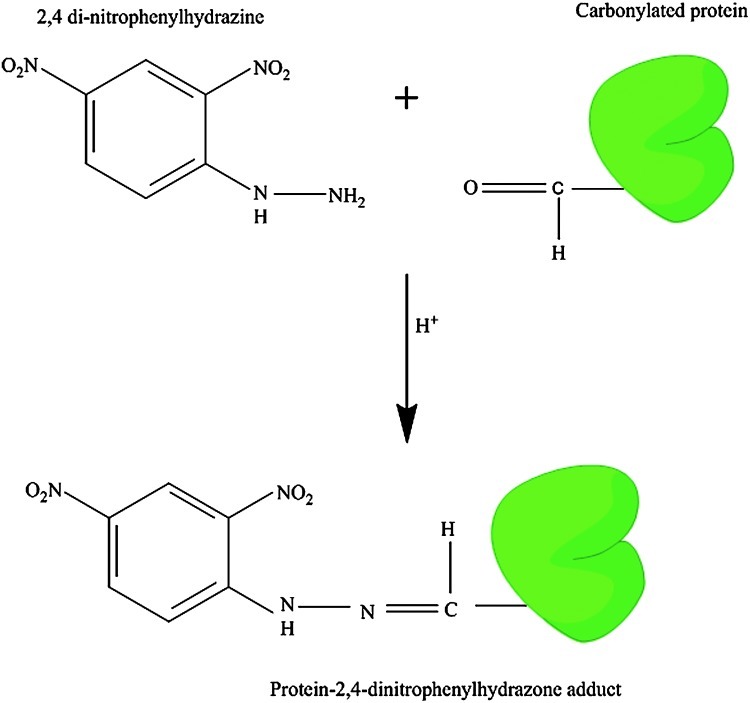
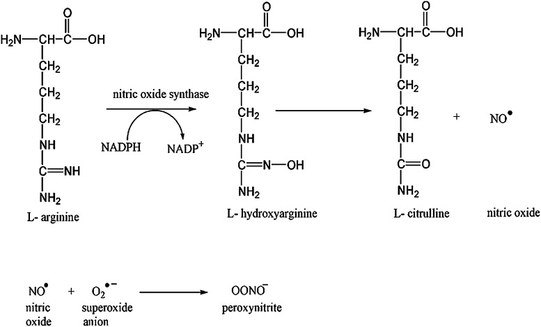
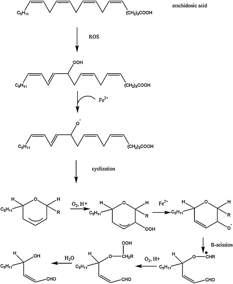
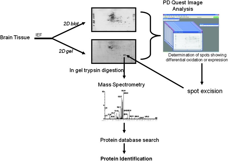
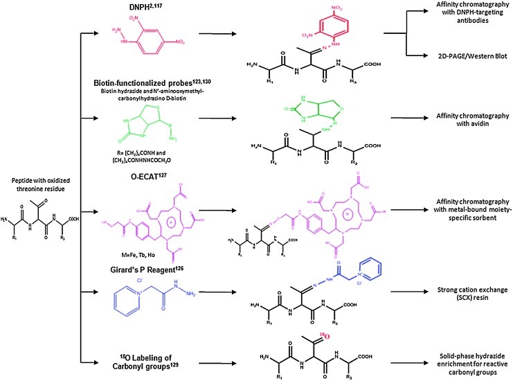
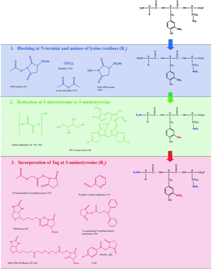
Several studies demonstrated that oxidative damage is a characteristic feature of many neurodegenerative diseases. The accumulation of oxidatively modified proteins may disrupt cellular functions by affecting protein expression, protein turnover, cell signaling, and induction of apoptosis and necrosis, suggesting that protein oxidation could have both physiological and pathological significance. For nearly two decades, our laboratory focused particular attention on studying oxidative damage of proteins and how their chemical modifications induced by reactive oxygen species/reactive nitrogen species correlate with pathology, biochemical alterations, and clinical presentations of Alzheimer's disease. This comprehensive article outlines basic knowledge of oxidative modification of proteins and lipids, followed by the principles of redox proteomics analysis, which also involve recent advances of mass spectrometry technology, and its application to selected age-related neurodegenerative diseases. Redox proteomics results obtained in different diseases and animal models thereof may provide new insights into the main mechanisms involved in the pathogenesis and progression of oxidative-stress-related neurodegenerative disorders. Redox proteomics can be considered a multifaceted approach that has the potential to provide insights into the molecular mechanisms of a disease, to find disease markers, as well as to identify potential targets for drug therapy. Considering the importance of a better understanding of the cause/effect of protein dysfunction in the pathogenesis and progression of neurodegenerative disorders, this article provides an overview of the intrinsic power of the redox proteomics approach together with the most significant results obtained by our laboratory and others during almost 10 years of research on neurodegenerative disorders since we initiated the field of redox proteomics.


Figures

FIG. 1.
FIG. 1.
Free radicals are generated by various mechanisms. One way by which free radicals are generated is via release of superoxide anion from the mitochondria, leading to increased formation of reactive oxygen and reactive nitrogen species and, consequently, damaging the biomolecules. (To see this illustration in color the reader is referred to the web version of this article at www.liebertonline.com/ars.)
FIG. 2.
FIG. 2.
Cysteine oxidation at neutral pH. Cysteine plays an important role in the regulation of protein function. Cysteine is vulnerable to attack by reactive oxygen species, which can lead to the formation of cysteine sufinic acid and eventually to the cysteine sulfonic acid. Measurement of the sulfonic acid on a protein is another maker for the detection of oxidative stress.
FIG. 3.
FIG. 3.
Derivatization of protein carbonyl using 2,4-dinitrophenylhydrazine (DNPH). The carbonyl group reacts with the DHPH to form a protein-DNPH hydrazone at acidic pH. This product is stable at neutral pH. The DNPH-protein hydrazone measures are used for the determination of the amount of oxidative damage to the protein in biological samples. (To see this illustration in color the reader is referred to the web version of this article at www.liebertonline.com/ars.)
FIG. 4.
FIG. 4.
Formation of peroxynitrite. During the conversion of L-arginine to L-Citrulline, nitric oxide is formed as one of the products. Nitric oxide can react with the superoxide anion, resulting in the formation of a highly reactive product, peroxynitrite.
FIG. 5.
FIG. 5.
The reaction of peroxynitrite and carbon dioxide results in the formation of nitrosoperoxylcarbonate, which undergoes rearrangement to form nitrocarbonate. Nitrocarbonate can undergo homolysis, resulting in the formation of nitrite radical and carbonate anion.
FIG. 6.
FIG. 6.
Formation of 3-NT from a tyrosine. The nitrite radical produced by the reaction of nitric oxide with carbon dioxide reacts with the tyrosine reside at meta-position, resulting in the formation of 3-NT. 3-NT, 3-nitrotyrosine.
FIG. 7.
FIG. 7.
One of the products of lipid peroxidation is HNE that can react with cysteine, lysine, and histine via Michael addition. Protein-bound HNE levels are used as an index of lipid peroxidation. HNE, 4-hydroxy-2-trans-nonenal.
FIG. 8.
FIG. 8.
Lipid peroxidation reaction summary. The process of lipid peroxidation involves an initiation process that begins with the hydrogen atom abstraction from an unsaturated fatty acid, resulting in the formation of lipid radical, which can then react with molecular oxygen, resulting in the formation of lipid peroxyl radicals. The lipid peroxyl radical can then abstract a H-atom from the other unsaturated fatty acid; this is referred to as a chain propagation reaction. When two lipid peroxyl radicals react, this will result in the termination of the lipid peroxidation process.
FIG. 9.
FIG. 9.
Formation of HNE from arachidonic acid. Oxidation of unsaturated fatty acids results in the formation of HNE.
FIG. 10.
FIG. 10.
Outline of redox proteomics approach. The identification of oxidatively modified protein involves first the separation of proteins by isoelectric point (IEF) followed by the separation of proteins based on relative mobility (Mr). The separation of the proteins is followed by transferring the proteins onto nitrocellulose or polyvinylidene fluoride membrane, probing with the antibody of interest, and determination of oxidatively modified protein by image analysis. Once a protein is identified as oxidatively modified, the protein spot will be excised from the gel, digested with trypsin, and subjected to mass spectrometry for correct identification of the proteins. (To see this illustration in color the reader is referred to the web version of this article at www.liebertonline.com/ars.)
FIG. 11.
FIG. 11.
Summary of methods for the derivatization and enrichment of carbonylated proteins are shown using an example tripeptide that contains an oxidized threonine residue. We note that other commonly carbonylated residues include Pro, Arg, Lys, His, and Trp, among others. (To see this illustration in color the reader is referred to the web version of this article at www.liebertonline.com/ars.)
FIG. 12.
FIG. 12.
Summary of strategies for the enrichment of 3-NT-modified proteins. In nongel based methods, 3-NT modified proteins were detected by blocking N-termini and amines of lysine residues. See text for references. (To see this illustration in color the reader is referred to the web version of this article at www.liebertonline.com/ars.)
FIG. 13.
FIG. 13.
Venn diagram of HNE-modified proteins identified during the progression of AD. Alpha-enolase and ATP synthase are the common targets of oxidative modification between AD, MCI, and EAD, and oxidative modification of these proteins might be key in the progression and pathogenesis of AD. AD, Alzheimer disease; EAD, early AD; MCI, mild cognitive impairment. (To see this illustration in color the reader is referred to the web version of this article at www.liebertonline.com/ars.)
FIG. 14.
FIG. 14.
Venn diagram of excessively carbonylated proteins throughout the pathogenesis of AD. Enolase, ATP synthase alpha, and UCH-L1 are the common targets of oxidation between AD, MCI, EAD, and PCAD. PCAD, preclinical Alzheimer disease; UCH-L1, ubiquitin carboxy-terminal hydrolase-L1. (To see this illustration in color the reader is referred to the web version of this article at www.liebertonline.com/ars.)
FIG. 15.
FIG. 15.
Nitrated proteins identified during the progression of AD in the hippocampus1 and IPL2 regions. DRP2 and aldolase are common targets of nitration between MCI and EAD. The identification of α-enolase as the only common target of protein nitration in AD, MCI, and EAD suggest that nitration of enolase might be critical to the progression and pathogenesis of AD. DRP2, dihydropyrimidinase-related protein 2; IPL, inferior parietal lobule. (To see this illustration in color the reader is referred to the web version of this article at www.liebertonline.com/ars.)
FIG. 16.
FIG. 16.
Redox proteomics determined functional pathways in different neurodegenerative disorders. A comparative analysis of the functional pathways involved in the redox proteomics-identified brain proteins from AD, PD, HD, and ALS showed that the proteins involved in glucose metabolism, mitochondrial function, cellular structure, and protein degradation are affected in common in these neurodenerative diseases. HD, Huntington disease; PD, Parkinson disease. (To see this illustration in color the reader is referred to the web version of this article at www.liebertonline.com/ars.)

References

    1. Abboud K. Bassila JC. Ghali-Ghoul R. Sabra R. Temporal changes in vascular reactivity in early diabetes mellitus in rats: role of changes in endothelial factors and in phosphodiesterase activity. Am J Physiol Heart Circ Physiol. 2009;297:H836–H845. - PubMed
    1. Abe K. Pan LH. Watanabe M. Kato T. Itoyama Y. Induction of nitrotyrosine-like immunoreactivity in the lower motor neuron of amyotrophic lateral sclerosis. Neurosci Lett. 1995;199:152–154. - PubMed
    1. Abe K. Pan LH. Watanabe M. Konno H. Kato T. Itoyama Y. Upregulation of protein-tyrosine nitration in the anterior horn cells of amyotrophic lateral sclerosis. Neurol Res. 1997;19:124–128. - PubMed
    1. Abello N. Barroso B. Kerstjens HA. Postma DS. Bischoff R. Chemical labeling and enrichment of nitrotyrosine-containing peptides. Talanta. 2010;80:1503–1512. - PubMed
    1. Adibhatla RM. Hatcher JF. Lipid oxidation and peroxidation in CNS health and disease: from molecular mechanisms to therapeutic opportunities. Antioxid Redox Signal. 2010;12:125–169. - PubMed

Publication types