. 2012 Mar;142(3):562-571.e2.
doi: 10.1053/j.gastro.2011.11.026. Epub 2011 Nov 22.

Smad4-mediated signaling inhibits intestinal neoplasia by inhibiting expression of β-catenin

Affiliations

Smad4-mediated signaling inhibits intestinal neoplasia by inhibiting expression of β-catenin

Tanner J Freeman et al. Gastroenterology. 2012 Mar.

Abstract

Background & aims: Mutational inactivation of adenomatous polyposis coli (APC) is an early event in colorectal cancer (CRC) progression that affects the stability and increases the activity of β-catenin, a mediator of Wnt signaling. Progression of CRC also involves inactivation of signaling via transforming growth factor β and bone morphogenetic protein (BMP), which are tumor suppressors. However, the interactions between these pathways are not clear. We investigated the effects of loss of the transcription factor Smad4 on levels of β-catenin messenger RNA (mRNA) and Wnt signaling.

Methods: We used microarray analysis to associate levels of Smad4 and β-catenin mRNA in colorectal tumor samples from 250 patients. We performed oligonucleotide-mediated knockdown of Smad4 in human embryonic kidney (HEK293T) and in HCT116 colon cancer cells and transgenically expressed Smad4 in SW480 colon cancer cells. We analyzed adenomas from (APC(Δ1638/+)) and (APC(Δ1638/+)) × (K19Cre(ERT2)Smad4(lox/lox)) mice by using laser capture microdissection.

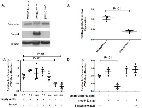
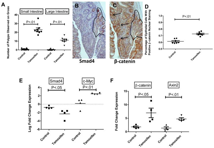
Results: In human CRC samples, reduced levels of Smad4 correlated with increased levels of β-catenin mRNA. In Smad4-depleted cell lines, levels of β-catenin mRNA and Wnt signaling increased. Inhibition of BMP or depletion of Smad4 in HEK293T cells increased binding of RNA polymerase II to the β-catenin gene. Expression of Smad4 in SW480 cells reduced Wnt signaling and levels of β-catenin mRNA. In mice with heterozygous disruption of Apc(APC(Δ1638/+)), Smad4-deficient intestinal adenomas had increased levels of β-catenin mRNA and expression of Wnt target genes compared with adenomas from APC(Δ1638/+) mice that expressed Smad4.

Conclusions: Transcription of β-catenin is inhibited by BMP signaling to Smad4. These findings provide important information about the interaction among transforming growth factor β, BMP, and Wnt signaling pathways in progression of CRC.


Figures

Figure 1
Figure 1. Smad4 depletion in cultured epithelial cells results in increased β-catenin expression and activation of TOPFlash activity
Oligonucleotide (RNAi) mediated Smad4 knockdown conducted in (A–C) HCT116 and (D–F) HEK293T human colon cancer cells. (A, D) Results of western blots showing relative expression of Smad4, Smad2, β-catenin and β-actin 24 hours post-transfection with scrambled or Smad4-specific RNAi (B, E) qPCR results showing relative expression of β-catenin mRNA expression 24hours post-transfection with scrambled and Smad4-specific RNAi. The graph shows quantified β-catenin mRNA relative to levels in mock-transfected cells normalized to Pmm1 expression. Triplicate for each condition was performed with mean +/−SEM displayed. Student’s t-tests were performed to determine significance. (C, F) TOPFlash activity 24 hours post-transfection with scrambled or Smad4 specific RNAi, with and without treatment with Wnt3a conditioned media. The graph shows light units from induced TOPFlash activity normalized to constitutive Renilla activity, relative to mock-transfected cells. Significance determined by ANOVA.
Figure 2
Figure 2. BMP signaling suppresses TOPFlash activity and regulates β-catenin mRNA expression levels
(A) SW480 cells were transiently co-transfected with either BRE-Luc and pCMV-Script or pCMV-Smad4 and then treated with either vehicle 20ng/ml BMP2 and/or 200ng/ml Noggin for 24 hours. Relative luciferase activity is graphed. Significance was determined by ANOVA tests. (B) Western blot of parallel experiments of HEK29T cells. Protein lysate was taken 30 min and 24hours after the indicated treatment to capture the early phosphorylation of R-Smads (p-Smad1, Smad1,p -Smad2, and Smad2) and the downstream effects of activation of the pathways (β-catenin and Id2). Representative β-actin taken at 24 hours is displayed. (C) HEK293T cells were treated with either vehicle, 200ng/ml BMP2, 5ng/ml TGF-β, or Wnt3a conditioned medium (1:1) and assayed for TOPflash activity at 24 hours post-transfection and 12 hours post-treatment. (D) Results of qPCR assays showing relative expression of β-catenin mRNA expression in HEK293T at 12 hours post-treatment with 20ng/ml BMP2, 200ng/ml Noggin or Wnt3a conditioned media. The graph shows quantified β-catenin mRNA relative to untreated cells. Duplicate for each condition was performed with mean +/−SEM displayed for all. Significance determined by student’s t-tests (C&D).
Figure 3
Figure 3. Smad4 restoration suppresses β-catenin mRNA expression and represses TOPFlash activity in a β-catenin dependent manner
((A) Representative results from three biological replicates of FACS sorted SW480 cells co-transfected with pRK-5Smad4 (0.8ug/mL) and pEGFP (1.0ug/mL). Protein isolated from GFP- (Smad4-null) and GFP+ (Smad4+) was run on western blot. (B) qPCR results from Smad4-null and Smad4+cells from (A) showing relative β-catenin expression that was normalized to Pmm1 expression. (C) TOPflash activity 48hrs post-transfection with p-RK5 empty vector or p-RK5-Smad4 vector as indicated. Triplicate for each condition were performed. (D) TOPFlash reporter activity in SW480 cells and in SW480 cells transiently co-transfected with 0.2μg wild-type human β-catenin and 0.8μg p-RK5 empty vector or 0.8μg p-RK5-Smad4 (as indicated). Two biological replicates are displayed. Significance determined using student’s t-test. Mean +/−SEM are displayed with FOP control (C&D).
Figure 4
Figure 4. BMP signaling regulates RNA polII activity of ctnnb1
HEK293T cells treated for 12 hours with BMP2, Noggin, or Wnt3a, as labeled, then nuclear lysates were prepared as described followed by immunoprecipitation with anti-RNA pol II antibody. (A) qPCR analysis of bound exon 2 of ctnnb1 using the LightCycler 480 System for analysis. (B) HEK293T cells transfected with either scrambled siRNA or Smad4 siRNA and after 24 hours were treated for 12 hours, as labeled. Nuclear lysates were prepared as described above, followed by immunoprecipitation with anti-RNA pol II antibody and amplification of exon 2 of ctnnb1 for qPCR analysis. Results presented are from two biological replicates run in triplicate.
Figure 5
Figure 5. Loss of Smad4 in vivo leads to increased tumor burden, levels of β-catenin mRNA, and downstream targets of Wnt Signaling
(A) Quantification of polyp burden observed on gross dissection in APCΔ1638/+K19CreERT2 Smad4lox/lox mice five weeks post-treatment (n=8 for both treatments). Serial sections (B, C) of APCΔ1638/+K19CreERT2 Smad4lox/lox polyp resected 5 weeks post-tamoxifen treatment. Photomicrographs were taken at 50x magnification. Scale bars equal 200μm (B) Smad4 immunostaining with outlined areas indicate corresponding Smad4-positive region. (C) β-catenin immunostaining with black arrows indicating areas of nuclear β-catenin staining. (D) Quantification of cells with positive nuclear β-catenin staining (defined as nuclear staining> cytoplasmic staining). Each data point represents the average percentage of positive cells within three high power fields for a single polyp (n=7 control polyps, n=7 tamoxifen polyps). Relative expression of (E) Smad4, c-Myc (F) β-catenin, Axin2 mRNA in vehicle (n=4) or tamoxifen (n=4) treated small bowel adenomas from APCΔ1638/+K19CreERT2Smad4lox/lox mice. Expression of mRNA is displayed as fold changes (2ΔCt) normalizing by the expression of target genes in normal adjacent tissue. Smad4 and c-Myc data are log transformed to display differences in a clear manner due to magnitude of difference. Significance determined by student’s t-tests.

References

    1. Clevers H. Wnt/beta-catenin signaling in development and disease. Cell. 2006 Nov 3;127(3):469–480. - PubMed
    1. Kosinski C, Li VS, Chan AS, et al. Gene expression patterns of human colon tops and basal crypts and BMP antagonists as intestinal stem cell niche factors. Proc Natl Acad Sci U S A. 2007 Sep 25;104(39):15418–15423. - PMC - PubMed
    1. Kinzler KW, Vogelstein B. Lessons from hereditary colorectal cancer. Cell. 1996 Oct 18;87(2):159–170. - PubMed
    1. Vermeulen L, De Sousa EMF, van der Heijden M, et al. Wnt activity defines colon cancer stem cells and is regulated by the microenvironment. Nat Cell Biol. May;12(5):468–476. - PubMed
    1. Brabletz S, Schmalhofer O, Brabletz T. Gastrointestinal stem cells in development and cancer. J Pathol. 2009 Jan;217(2):307–317. - PubMed

Publication types

MeSH terms

Associated data