. 2012 Feb;45(2):774-85.
doi: 10.1016/j.nbd.2011.11.001. Epub 2011 Nov 13.

Interictal spikes, seizures and ictal cell death are not necessary for post-traumatic epileptogenesis in vitro

Affiliations

Interictal spikes, seizures and ictal cell death are not necessary for post-traumatic epileptogenesis in vitro

Yevgeny Berdichevsky et al. Neurobiol Dis. 2012 Feb.

Abstract

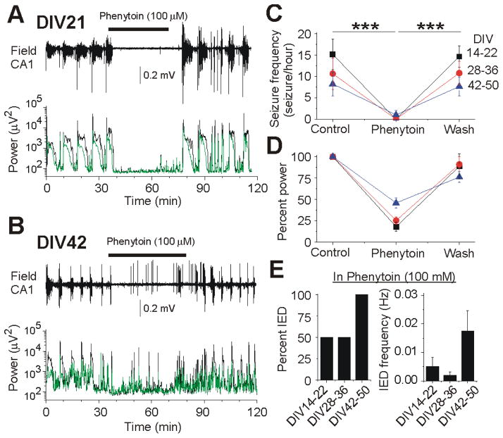
Clinical studies indicate that phenytoin prevents acute post-traumatic seizures but not subsequent post-traumatic epilepsy. We explored this phenomenon using organotypic hippocampal slice cultures as a model of severe traumatic brain injury. Hippocampal slices were cultured for up to eight weeks, during which acute and chronic electrical recordings revealed a characteristic evolution of spontaneous epileptiform discharges, including interictal spikes, seizure activity and electrical status epilepticus. Cell death exhibited an early peak immediately following slicing, and a later secondary peak that coincided with the peak of seizure-like activity. The secondary peak in neuronal death was abolished by either blockade of glutamatergic transmission with kynurenic acid or by elimination of ictal activity and status epilepticus with phenytoin. Withdrawal of kynurenic acid or phenytoin was followed by a sharp increase in spontaneous seizure activity. Phenytoin's anticonvulsant and neuroprotective effects failed after four weeks of continuous administration. These data support the clinical findings that after brain injury, anticonvulsants prevent seizures but not epilepsy or the development of anticonvulsant resistance. We extend the clinical data by showing that secondary neuronal death is correlated with ictal but not interictal activity, and that blocking all three of these sequelae of brain injury does not prevent epileptogenesis in this in vitro model.


Figures

Fig. 1
Fig. 1
Chronic electrical recording in rat organotypic hippocampal slices in vitro. (A), schematic of the custom MEA with four organotypic hippocampal cultures. (B), micrograph of one of the hippocampal cultures on MEA, scale bar is 200 μm. (C), raster plot of electrical activity recorded in an organotypic culture. (D), the types of activity detected with an electrode (top), and the color map of interictal activity (top trace), and ictal activity (bottom trace), each recording is 1 hour long. Arrows point to interictal spikes and ictal events shown at different time scale, trace duration is 100 seconds. (E), Incidence of ictal and interictal activity as a percentage of cultures recorded on MEAs, with age of cuture. (F), Cumulative duration of ictal activity observed per culture with age. N = 7 cultures for (E) and (F).
Fig. 2
Fig. 2
Epileptogenesis in mouse organotypic hippocampal slices. (A–B) Extracellular field potential recordings in the CA3 pyramidal cell layer in organotypic hippocampal slices at DIV 7 (A) and DIV14 (B). Electrical recordings revealed spontaneous epileptiform discharges, including interictal epileptiform discharges (IEDs) and ictal-like seizure activity. (C) Group data from 70 recordings (2–3 hours in each recording) from 70 organotypic hippocampal slices (n = 10 slices in each age group).
Fig. 3
Fig. 3
Activity-dependent cell death in post-traumatic epileptogenesis. (A) Time course of cell death in epileptic rat organotypic cultures. Propidium iodide uptake is a marker of cell death. Initial high counts of PI-positive cells reflect high number of dead cells following trauma (dissection). Cell death drops during the latent period, and then increases as control cultures transition from interictal to ictal activity. KYNA-treated cultures do not have epileptiform activity, and largely avoid second wave of cell death (** represents p < 0.01, *** p < 0.001, n = 6 slices in control and KYNA groups). (B) schematic representation of progression of epilepsy in organotypic cultures. (C) number of dead (PI-positive) neurons in CA1 pyramidal layer (CA1-pcl) and in the dentate gyrus granule cell layer (DG gcl) is much higher in control mouse cultures than cultures where synchronous activity has been blocked with kynurenic acid (KYNA). (D) top, evolution of cell death with time in CA1 pcl, bottom, in DG gcl in cultures (n = 7 control mouse slices, n = 5 KYNA-treated mouse slices, * represents p < 0.05. Error bars show standard deviation). (E, F) neuron density in CA1 pyramidal layer is significantly higher in chronically KYNA-treated mouse cultures than in controls at 21 DIV, standard deviation shown as error bars. (G, H) ictal-like tonic-clonic electrical seizures resume after washout of KYNA from chronically treated cultures, showing lack of effect on epileptogenesis (n = 8 cultures).
Fig. 4
Fig. 4
Concentration dependent efficacy of Phenytoin in post-traumatic seizures. (A–B) Extracellular field potential recordings from cultured mouse hippocampal slices at DIV21 and DIV20 before (control), during and after (wash) applications of 30 μM (A) and 100 μM (B) phenytoin. Plots below show corresponding power of spontaneous electrical activity. (C–D) Mean frequency of spontaneous seizure activity and power of electrical activity before, during and after phenytoin (30 and 100 μM) application. (E) Group data of the spontaneous interictal discharges (IED) in phenytoin (30 μM) and phenytoin (100 μM) (n = 6 for each group).
Fig. 5
Fig. 5
Acute administration of phenytoin suppresses seizures at all stages of epileptogenesis. (A–B) Extracellular field potential recordings from cultured mouse hippocampal slices at DIV21 and DIV42. Plots below show power of electrical activity in the CA3 and CA1 pyramidal cell layer before (control), during and after phenytoin (100 μM) application. (A) A complete anticonvulsant response to acute phenytoin including suppression of all ictal and interictal activity. Application of phenytoin abolished spontaneous interictal bursts and recurrent seizures and strongly reduced power amplitude in EEG (1–100 Hz) and fast ripple (100–500 Hz) frequency range. (B) Example of a partial anticonvulsant effect of phenytoin. Application of Phenytoin abolished recurrent seizures, but not interictal epileptiform discharges, and so only partially reduced the power amplitude. (C) Fraction of hippocampal slices in which phenytoin achieved full and partial effects on spontaneous epileptiform discharges. (D) Mean power of spontaneous electrical activity (percent of control) in EEG and fast ripple frequency range after acute application of phenytoin at each stage of epileptogenesis. (E) Summary of phenytoin effects on interictal activity at each stage.
Fig. 6
Fig. 6
Loss of anticonvulsant effect of phenytoin after chronic exposure. (A) Extracellular field potential recording from organotypic mouse hippocampal slice at DIV 20. Plot below shows corresponding power of electrical activity in 8.2 s windows. Removal of Phenytoin from incubation medium resulted was followed by a sharp increase in seizure activity. (B) Extracellular field potential recording and corresponding power of electrical activity in organotypic hippocampal slice at DIV42. Spontaneous seizures are marked by asterisks. (C–D) Frequency of seizures and power of extracellular field potential activity in the individual recordings. (E) Mean seizure frequency and changes of power of electrical activity. After four-five weeks, chronic incubation with phenytoin was less effective at controlling seizures.
Fig. 7
Fig. 7
Loss of neuroprotective effect of chronic phenytoin administration. Mouse slice cultures chronically exposed to 100 μM phenytoin from 3DIV. At 14 DIV, phenytoin significantly reduces neuronal death in CA1 pyramidal layer. At 36 DIV, when phenytoin loses efficacy as an anticonvulsant, cell death in cultures chronically treated with phenytoin is not significantly different from controls (n = 5 slices, each condition, error bars show standard deviation, * represents p < 0.05)

References

    1. Annegers JF, Hauser WA, Coan SP, Rocca WA. A population-based study of seizures after traumatic brain injuries. N Engl J Med. 1998;338:20–4. - PubMed
    1. Ben-Ari Y, Dudek FE. Primary and secondary mechanisms of epileptogenesis in the temporal lobe: there is a before and an after. Epilepsy Curr. 2010;10:118–25. - PMC - PubMed
    1. Berdichevsky Y, Sabolek H, Levine JB, Staley KJ, Yarmush ML. Microfluidics and multielectrode array-compatible organotypic slice culture method. J Neurosci Methods 2008 - PMC - PubMed
    1. Berg AT. The natural history of mesial temporal lobe epilepsy. Curr Opin Neurol. 2008;21:173–8. - PubMed
    1. Bernhardt BC, Worsley KJ, Kim H, Evans AC, Bernasconi A, Bernasconi N. Longitudinal and cross-sectional analysis of atrophy in pharmacoresistant temporal lobe epilepsy. Neurology. 2009;72:1747–54. - PMC - PubMed

Publication types

MeSH terms