

Assessing abnormal iron content in the deep gray matter of patients with multiple sclerosis versus healthy controls
- PMID: 22116106
- PMCID: PMC7964804
- DOI: 10.3174/ajnr.A2773
Assessing abnormal iron content in the deep gray matter of patients with multiple sclerosis versus healthy controls
Abstract
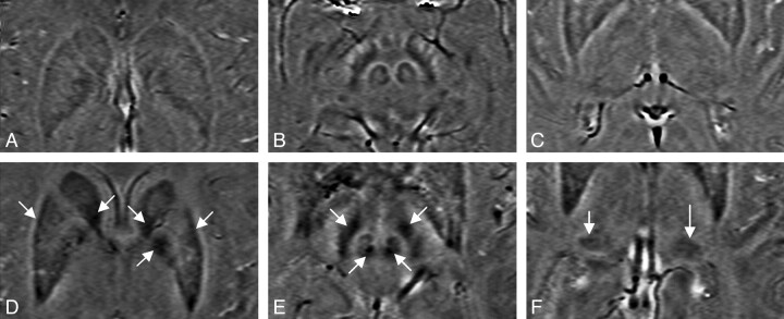
Background and purpose: It is well known that patients with MS tend to have abnormal iron deposition in and around the MS plaques, in the basal ganglia and the THA. In this study, we used SWI to quantify iron content in patients with MS and healthy volunteers.
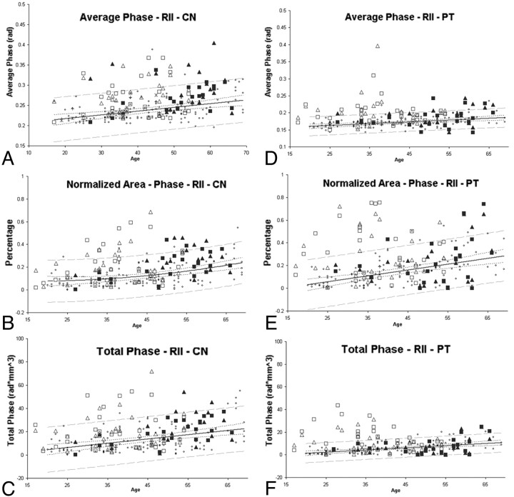
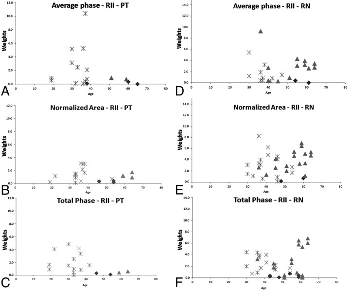
Materials and methods: Fifty-two patients with MS were recruited to assess abnormal iron content in their basal ganglia and THA structures. One hundred twenty-two healthy subjects were recruited to establish a baseline of normal iron content in deep GM structures. Each structure was separated into 2 regions: a low-iron-content region and a high-iron-content region. The average phase, the percentage area, and the total phase of the high-iron-content region were evaluated. A weighting was also assigned to each subject depending on the level of iron content and its deviation from the normal range.
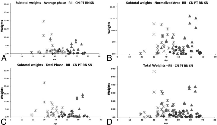
Results: A clear separation between iron content in healthy subjects versus patients with MS was seen. For healthy subjects 13% and for patients with MS 65% showed an iron-weighting factor >3 SDs from the normal mean (P < .05). The results for those patients younger than 40 years are even more impressive. In these cases, only 1% of healthy subjects and 67% of patients with RRMS showed abnormally high iron content.
Conclusions: Iron-weighting factors in the basal ganglia, THA, and the midbrain appeared to be abnormal in roughly two-thirds of patients with MS as measured by SWI.
Figures
Similar articles
-
Iron stores and cerebral veins in MS studied by susceptibility weighted imaging.Int Angiol. 2010 Apr;29(2):149-57. Int Angiol. 2010. PMID: 20351671
-
Iron deposition in the gray matter in patients with relapse-remitting multiple sclerosis: A longitudinal study using three-dimensional (3D)-enhanced T2*-weighted angiography (ESWAN).Eur J Radiol. 2015 Jul;84(7):1325-32. doi: 10.1016/j.ejrad.2015.04.013. Epub 2015 Apr 25. Eur J Radiol. 2015. PMID: 25959392
-
Quantitative assessment of iron accumulation in the deep gray matter of multiple sclerosis by magnetic field correlation imaging.AJNR Am J Neuroradiol. 2007 Oct;28(9):1639-44. doi: 10.3174/ajnr.A0646. Epub 2007 Sep 24. AJNR Am J Neuroradiol. 2007. PMID: 17893225 Free PMC article.
-
Magnetic resonance imaging signatures of vascular pathology in multiple sclerosis.Neurol Res. 2012 Oct;34(8):780-92. doi: 10.1179/1743132812Y.0000000078. Neurol Res. 2012. PMID: 22971468 Review.
-
Iron Mapping in Multiple Sclerosis.Neuroimaging Clin N Am. 2017 May;27(2):335-342. doi: 10.1016/j.nic.2016.12.003. Epub 2017 Jan 20. Neuroimaging Clin N Am. 2017. PMID: 28391790 Review.
Cited by
-
Quantitative Susceptibility Mapping Values Quantification in Deep Gray Matter Structures for Relapsing-Remitting Multiple Sclerosis: A Systematic Review and Meta-Analysis.Brain Behav. 2024 Oct;14(10):e70093. doi: 10.1002/brb3.70093. Brain Behav. 2024. PMID: 39415615 Free PMC article.
-
Iron Content in Deep Gray Matter as a Function of Age Using Quantitative Susceptibility Mapping: A Multicenter Study.Front Neurosci. 2021 Jan 6;14:607705. doi: 10.3389/fnins.2020.607705. eCollection 2020. Front Neurosci. 2021. PMID: 33488350 Free PMC article.
-
Brain Iron at Quantitative MRI Is Associated with Disability in Multiple Sclerosis.Radiology. 2018 Nov;289(2):487-496. doi: 10.1148/radiol.2018180136. Epub 2018 Jul 17. Radiology. 2018. PMID: 30015589 Free PMC article.
-
Mapping of thalamic magnetic susceptibility in multiple sclerosis indicates decreasing iron with disease duration: A proposed mechanistic relationship between inflammation and oligodendrocyte vitality.Neuroimage. 2018 Feb 15;167:438-452. doi: 10.1016/j.neuroimage.2017.10.063. Epub 2017 Oct 31. Neuroimage. 2018. PMID: 29097315 Free PMC article.
-
Evaluation of iron deposition in brain basal ganglia of patients with Parkinson's disease using quantitative susceptibility mapping.Eur J Radiol Open. 2019 Apr 29;6:169-174. doi: 10.1016/j.ejro.2019.04.005. eCollection 2019. Eur J Radiol Open. 2019. PMID: 31065578 Free PMC article.
References
-
- Schelling F. Damaging venous reflux into the skull or spine: relevance to multiple sclerosis. Med Hypotheses 1986; 21: 141– 48 - PubMed
-
- Singh AV, Zamboni P. Anomalous venous blood flow and iron deposition in multiple sclerosis. J Cereb Blood Flow Metab 2009; 29: 1867– 78. Epub 2009 Sep 2 - PubMed
-
- Filippi M, Rocca MA, Martino G, et al. . Magnetization transfer changes in the normal appearing white matter precede the appearance of enhancing lesions in patients with multiple sclerosis. Ann Neurol 1998; 43: 809– 14 - PubMed
Publication types
MeSH terms
Substances
LinkOut - more resources
Full Text Sources
Medical