. 2012 Mar;33(3):513-8.
doi: 10.3174/ajnr.A2814. Epub 2011 Nov 24.

Asymmetric development of the hippocampal region is common: a fetal MR imaging study

Affiliations

Asymmetric development of the hippocampal region is common: a fetal MR imaging study

D Bajic et al. AJNR Am J Neuroradiol. 2012 Mar.

Abstract

Background and purpose: Hippocampal development is poorly understood. This study evaluated the normal development of the hippocampal region during the fetal period by using MR imaging.

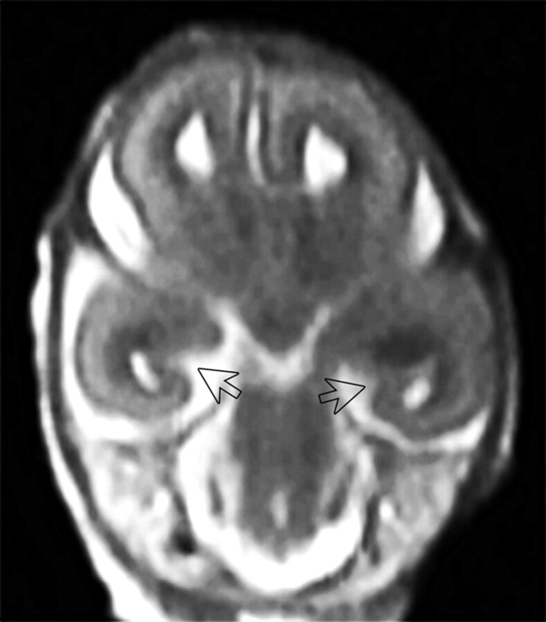
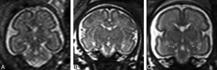
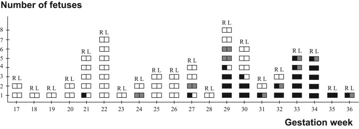
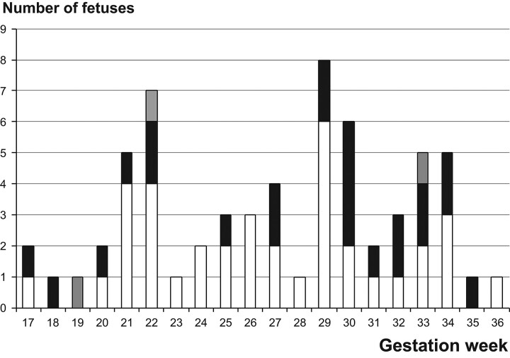
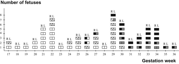
Materials and methods: MR images of 63 fetuses without intracranial pathology were reviewed independently by 2 radiologists with no knowledge of the fetal GA. Three MR images were performed postmortem and 60 in vivo. The progress of hippocampal inversion was analyzed in coronal sections, and the left and right sides of the hippocampal region were compared in every case.

Results: The fetuses in the postmortem examinations were at GWs 17-18 and in the in vivo examinations, at GWs 19-36. The hippocampal sulcus was open, bi- or unilaterally, in 39 fetuses. The oldest was at GW 32. The sulcus was closed at GW 21 at the earliest, unilaterally. In 26/63 fetuses (41%), the deepening or closure of the hippocampal sulcus or hippocampal inversion was asymmetric; in 23 fetuses, the right side developed faster. A shallow collateral sulcus was found earliest at GW 17. A deep collateral sulcus was visible earliest at GW 26 unilaterally, but in all fetuses from GW 31 onward, it was seen bilaterally. The orientation of the collateral sulcus was not related to the GA.

Conclusions: There are wide individual temporal variations in the development and the inversion process of the hippocampal sulcus as well as in the formation of the collateral sulcus. Asymmetric development is common, and in most of the asymmetric cases, the right hippocampus develops faster.


Figures

Fig 1.
Fig 1.
Postmortem MRI study of a fetus aborted at 17 GW. The arrows indicate widely open symmetric hippocampal sulci. T2-weighted TSE coronal image.
Fig 2.
Fig 2.
In vivo T2-weighted single-shot FSE coronal images. A, Fetus at GW 25. The arrows indicate the hippocampal sulci. The hippocampal sulcus is larger on the left side. B, Fetus at GW 29. Closed hippocampal sulcus and a nonoval hippocampus with a vertical long axis bilaterally. The arrows indicate the hippocampal region, and the stars, the collateral sulcus. C, Fetus at GW 29. Closed hippocampal sulcus and an oval hippocampus with a horizontal long axis bilaterally. White arrows indicate the hippocampal region; the black arrows, the closed hippocampal sulcus; and the star, the collateral sulcus.
Fig 3.
Fig 3.
Closure of the hippocampal sulcus and the shapes of the hippocampi in fetuses with a closed hippocampal sulcus. Each square pair describes a fetus. R indicates right side; L, left side; open square, open hippocampal sulcus; shaded square, nonoval shape of the inverted hippocampus; black square, oval shape of the inverted hippocampus, with the long axis horizontal (complete inversion).
Fig 4.
Fig 4.
Comparison of the development of the left and right sides of the hippocampus. Open box indicates symmetric development; shaded square, the left side more developed; black square, the right side more developed.
Fig 5.
Fig 5.
Collateral sulcus at different GAs. Each square pair describes a fetus. R indicates right side; L, left side. Collateral sulcus: Open square indicates not visible; slashed square, shallow; shaded square, vertical; black square, oblique or horizontal.

References

    1. Kier EL, Fulbright RK, Bronen RA. Limbic lobe embryology and anatomy: dissection and MR of the medial surface of the fetal cerebral hemisphere. AJNR Am J Neuroradiol 1995; 16: 1847–53 - PMC - PubMed
    1. Kier EL, Kim JH, Fulbright RK, et al. . Embryology of the human fetal hippocampus: MR imaging, anatomy, and histology. AJNR Am J Neuroradiol 1997; 18: 525–32 - PMC - PubMed
    1. Rados M, Judas M, Kostovic I. In vitro MRI of brain development. Eur J Radiol 2006; 57: 187–98 - PubMed
    1. Humphrey T. The development of the human hippocampal fissure. J Anat 1967; 101: 655–76 - PMC - PubMed
    1. Arnold SE, Trojanowski QJ. Human fetal hippocampal development, I. Cytoarchitecture, myeloarchitecture and neuronal morphologic features. J Comp Neurol 1996; 367: 274–92 - PubMed