

Role of ultrasound in the assessment of benignity and malignancy of parotid masses
- PMID: 22116132
- PMCID: PMC3520365
- DOI: 10.1259/dmfr/60907848
Role of ultrasound in the assessment of benignity and malignancy of parotid masses
Abstract
Objectives: This study aimed to investigate the value of ultrasound in the identification of benign and malignant parotid masses.
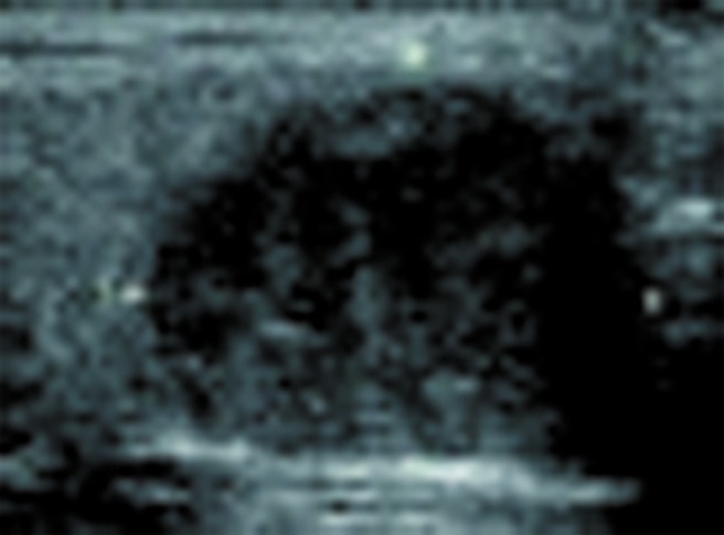
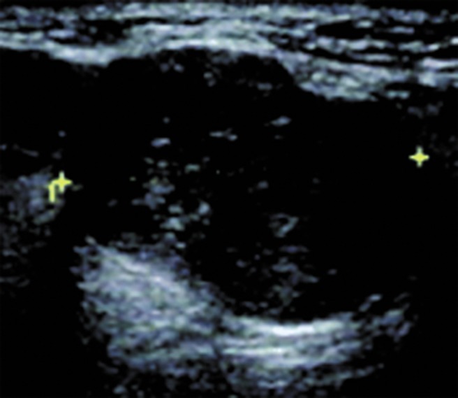
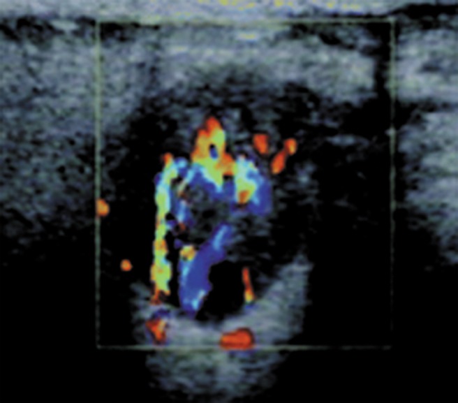
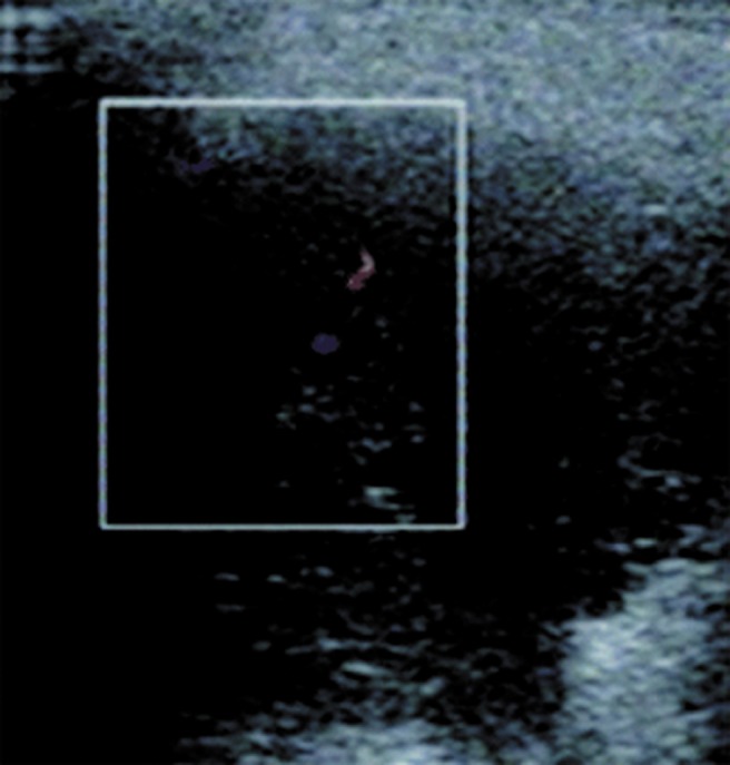
Methods: Data of 189 patients with parotid gland masses undergoing ultrasound-guided fine-needle aspiration (FNA), core biopsy or surgery were reviewed retrospectively and the presumed sonographic diagnoses were compared with the histopathology. The sensitivity, specificity and accuracy of sonographic diagnoses were assessed and the sonographic characteristics of those lesions, including shape, margin, echogenicity, echotexture and vascularization, were studied.
Results: Of the 189 patients, the final pathological diagnosis included 18 malignant tumours and 171 benign masses; the presumed sonographic diagnoses showed 165 cases as benign and probably benign masses (11 cases were confirmed malignant, 154 cases benign) and 24 cases were diagnosed as probably malignant and malignant masses (7 cases were confirmed malignant, 17 cases benign). The sensitivity, specificity, positive predictive value, negative predictive value and accuracy of ultrasound for the diagnosis of parotid gland masses were 38.9%, 90.1%, 29.2%, 93.3% and 85.2%, respectively, and accuracy for malignant masses was 20%. The sonographic characteristics of parotid masses between benign and malignant lesions had no significant differences. The parotid gland masses in this study included pleomorphic adenoma, Warthin's tumour, retention cyst, haemangiomas, chronic granuloma, lymphoma, fibrolipoma, abscess, basal cell adenoma, oncocytoma, lymphatic tuberculosis, myoepithelioma, neurilemmoma, mucoepidermoid carcinoma, adenoid cystic carcinoma, alveolar soft part sarcoma and retinal blastoma (metastasis).
Conclusions: It is challenging to use sonography for differentiating between benign and malignant parotid gland masses. To make a definite diagnosis, ultrasound-guided FNA or core biopsy is advocated.
Figures

Similar articles
-
Clinicopathological analysis of parotid masses: six-year experience of a tertiary center.J Pak Med Assoc. 2020 Feb;70(2):308-312. doi: 10.5455/JPMA.17185. J Pak Med Assoc. 2020. PMID: 32063626
-
Parotid tumors: differentiation of benign and malignant tumors with quantitative sonographic analyses.Ultrasound Med Biol. 2004 May;30(5):567-74. doi: 10.1016/j.ultrasmedbio.2004.02.007. Ultrasound Med Biol. 2004. PMID: 15183220
-
[Sensitivity and specificity of fine needle aspiration biopsy in parotid masses].Kulak Burun Bogaz Ihtis Derg. 2007;17(2):96-9. Kulak Burun Bogaz Ihtis Derg. 2007. PMID: 17527061 Turkish.
-
Gray scale and Doppler ultrasonography of the benign tumors of parotid gland (pleomorphic adenoma and Warthin's tumor). Pictorial essay.Med Ultrason. 2010 Sep;12(3):238-44. Med Ultrason. 2010. PMID: 21203603 Review.
-
Synchronous benign and malignant salivary gland tumors in ipsilateral glands: a report of two cases and a review of literature.Head Neck. 2002 Mar;24(3):301-6. doi: 10.1002/hed.10048. Head Neck. 2002. PMID: 11891964 Review.
Cited by
-
A Retrospective 8-Year Single Institutional Study in Germany Regarding Diagnosis, Treatment, and Outcome of Malignant Parotid Tumors.Int J Surg Oncol. 2024 Dec 9;2024:7598063. doi: 10.1155/ijso/7598063. eCollection 2024. Int J Surg Oncol. 2024. PMID: 39687544 Free PMC article.
-
Performance Evaluation of Ultrasound Images Using Non-Local Means Algorithm with Adaptive Isotropic Search Window for Improved Detection of Salivary Gland Diseases: A Pilot Study.Diagnostics (Basel). 2024 Jul 4;14(13):1433. doi: 10.3390/diagnostics14131433. Diagnostics (Basel). 2024. PMID: 39001323 Free PMC article.
-
Nodular fasciitis. A rare, rapidly growing lesion of the parotid gland.Intern Emerg Med. 2025 Jun;20(4):1271-1272. doi: 10.1007/s11739-025-03868-9. Epub 2025 Feb 1. Intern Emerg Med. 2025. PMID: 39891820 No abstract available.
-
Characteristic power Doppler sonographic images of tumorous and non-tumorous buccal space lesions.Dentomaxillofac Radiol. 2013;42(7):20120460. doi: 10.1259/dmfr.20120460. Epub 2013 Mar 21. Dentomaxillofac Radiol. 2013. PMID: 23520393 Free PMC article.
-
The Diagnostic Value of Ultrasound-Based Deep Learning in Differentiating Parotid Gland Tumors.J Oncol. 2022 May 12;2022:8192999. doi: 10.1155/2022/8192999. eCollection 2022. J Oncol. 2022. PMID: 35602298 Free PMC article.
References
-
- Białek EJ, Jakubowski W, Karpińska G. Role of ultrasonography in diagnosis and differentiation of pleomorphic adenomas. Arch Otolaryngol Head Neck Surg 2003;129:929–933 - PubMed
-
- Kim J, Kim EK, Park CS, Choi YS, Kim YH, Choi EC. Characteristic sonographic findings of Warthin's tumor in the parotid gland. J Clin Ultrasound 2004;32:78–81 - PubMed
-
- Howlett DC. High resolution ultrasound assessment of the parotid gland. Br J Radiol 2003;76:271–277 - PubMed
-
- Yonetsu K, Ohki M, Kumazawa S, Eida S, Sumi M, Nakamura T. Parotid tumors: differentiation of benign and malignant tumors with quantitative sonographic analyses. Ultrasound Med Biol 2004;30:567–574 - PubMed
-
- Bozzato A, Zenk J, Greess H, Hornung J, Gottwald F, Rabe C, et al. Potential of ultrasound diagnosis for parotid tumors: analysis of qualitative and quantitative parameters. Otolaryngol Head Neck Surg 2007;137:642–646 - PubMed
