

Branchial cleft cyst at an unusual location: a rare case with a brief review
- PMID: 22116133
- PMCID: PMC3528200
- DOI: 10.1259/dmfr/59515421
Branchial cleft cyst at an unusual location: a rare case with a brief review
Abstract
A branchial cleft cyst (BCC) commonly presents as a solitary, painless mass in the neck of a child or young adult. They are most commonly located along the anterior border and the upper third of the sternocleidomastoid muscle in the anterior triangle of the neck. It is very rare for a BCC to manifest in other locations, especially in the posterior triangle of the neck. BCCs are believed to be derived from the branchial apparatus, mostly from the second branchial arch, although many theories have been proposed to explain the aetiology of BCCs. It is possible for BCCs to be easily misdiagnosed as other swellings of oral or paraoral origin owing to their location. Intraoral lymphoepithelial cysts have also been reported in the literature. It is imperative that clinicians make an accurate diagnosis so that appropriate treatment can be performed. If the cysts are excised properly, recurrence is rare. A rare case report of BCC arising in the neck from an unusual location with components in the posterior triangle is presented here.
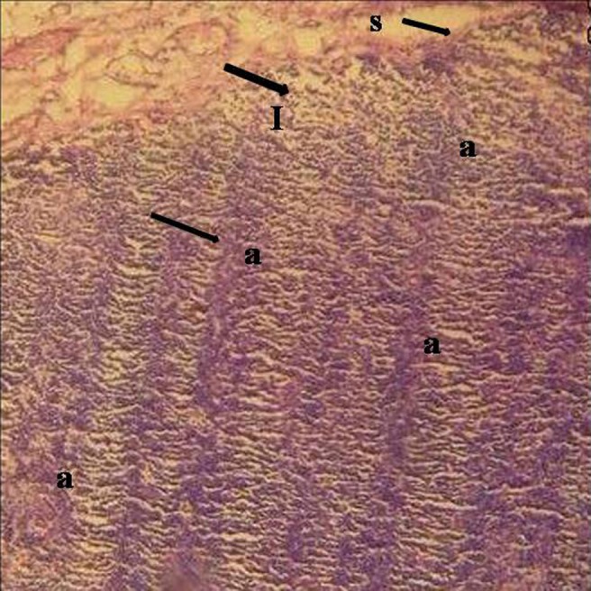
Figures


References
-
- Thomaidis V, Seretis K, Tamiolakis D, Papadopoulos N, Tsamis I. Branchial cysts—a report of 4 cases. Acta Dermatoven APA 2006;15:85–89 - PubMed
-
- Berseth CL, Poenaru D. Structural anomalies of GIT. In: Tacusch HW, Ballard RA, Gleason CA, eds. Avery’s diseases of the newborn (8th edn). Philadelphia, PA: WB Saunders, 2005. pp. 1086–1087
-
- Howard DJ, Lund VJ. Pharynx, larynx and neck—branchial cleft cyst. In: NS Williams, CJK Butstrode, PR O’Connell, A Hodder, eds. Bailey and Love’s short practise of surgery (20th edn). London, UK: Harcourt, 2008. pp. 727–729
-
- Glosser JW, Pires CA, Feinberg SE. Branchial cleft or cervical lymphoepithelial cysts: etiology and management. J Am Dent Assoc 2003;134:81–86 - PubMed
-
- Vaidya S, Pagare RS, Sharma VK. Lateral cervical cyst. Inter J Otorhinolaryngol 2008;7:1–6
Publication types
MeSH terms
Substances
LinkOut - more resources
Full Text Sources
Medical
