. 2012 Feb;56(2):179-87.
doi: 10.1007/s10493-011-9508-7. Epub 2011 Nov 25.

Life cycle, growth characteristics and host cell response of Rickettsia helvetica in a Vero cell line

Affiliations

Life cycle, growth characteristics and host cell response of Rickettsia helvetica in a Vero cell line

Karin Elfving et al. Exp Appl Acarol. 2012 Feb.

Erratum in

  • Exp Appl Acarol. 2012 Feb;56(2):189-90

Abstract

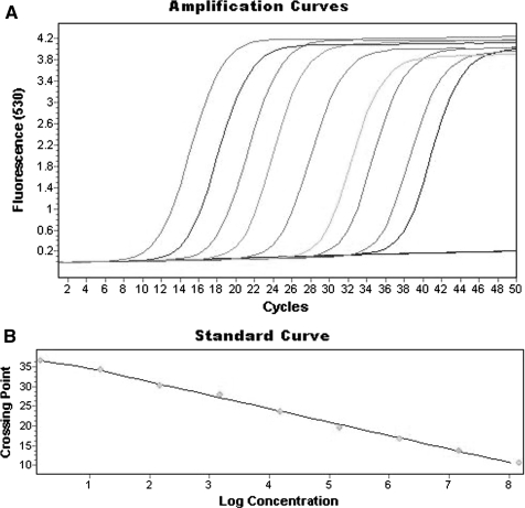
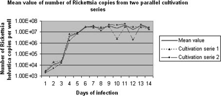
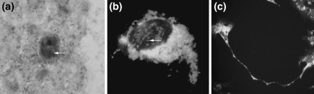
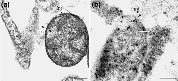
Rickettsia helvetica, a spotted fever rickettsia and emerging pathogen with Ixodes ricinus ticks as the main vector, is an agent of human disease and may cause febrile illness as well as meningitis. In three parallel series the isolated standard type of R. helvetica, obtained from a PCR-positive I. ricinus tick, was high-passaged and propagated in a Vero cell line. By using quantitative real-time PCR, the generation time from inoculation to stationary phase of growth was calculated to 20-22 h. In the static cultivation system the stationary phase was observed from the seventh day after inoculation, and there was no observed degradation of R. helvetica DNA during the 14 days studied. Microscopy showed that the organisms invaded the host cells rapidly and were primarily found free in the cytoplasm and only occasionally located in the nucleus. Four days after inoculation some of the host cells were broken and many indifferent stages of cytoplasmic organic decomposition were seen. However the R. helvetica organism did not show any morphologic alterations and the number of organisms was stable after the replication peak which may indicate that R. helvetica is adapted to growth in a Vero cell line and/or that the phase of degradation occurs later than the 14 days studied. The findings differ from what has been reported for other rickettsiae of the spotted fever group and may be of importance for invasiveness and virulence of R. helvetica.


Figures

Fig. 1
Fig. 1
a qPCR amplification in a dilution series of the plasmid pCR4-TOPO cloning vector containing the inserted gltA fragment of Rickettsia helvetica (standard type). b Standard calibration curve for R. helvetica plasmid pCR4-TOPO cloning vector showing linearity in the dilution series between 1.5 and 1.5 × 108 copies (Error 0.0142, Efficiency 1.961, R2 0.997, y=−3.34x+37.68)
Fig. 2
Fig. 2
qPCR of content from each well with actual and mean values of the number of Rickettsia helvetica copies in two parallel cultivation series in Vero cells for 14 days
Fig. 3
Fig. 3
Fourth day of Rickettsia helvetica infected Vero cells. a Vero cell with Gimenez stained rickettsiae in the cytoplasm. Single bacteria can be seen in the nucleus (arrow) ×400. b Fluorescent photomicrograph of infected Vero cell where a sparse number of bacteria are seen in the nucleus ×1000. c Fluorescent photomicrograph of a filopodia filled with fluorescent coccobacillary rickettsiae stained with specific rabbit anti-rickettsia antibodies ×200
Fig. 4
Fig. 4
Fourth to fifth day of Rickettsia helvetica infected Vero cells. a Morphology of R. helvetica in partly decomposed host cells. Note the leaflets (arrow heads) and inner plasma membrane enclosing the periplasmatic space (ps). Fibrillate nucleic acid is clearly visible (long arrows) ×120,000. Bar  150 nm. b Anti-rickettsia antibodies with gold particles (gp) (15 nm) on Lowicryl-embedded cells. Leaflets and plasma membrane are visible (arrows heads) as well as fibrillar nucleic acid (long arrows). The immunoreaction is mainly located along the membrane/leaflet part of the rickettsia but sparsely scattered all over the organism ×120,000. Bar  150 nm

References

    1. Balraj P, Vestris D, Raoult D, Renesto P. Comparison of Rickettsia conorii growth within different cell lines by real-time quantitative PCR. Clin Microbiol Infect. 2009;15:294–295. doi: 10.1111/j.1469-0691.2008.02243.x. - DOI - PubMed
    1. Beati L, Peter O, Burgdorfer W, Aeschlimann A, Raoult A. Confirmation that Rickettsia helvetica sp. nov. is a distinct species of the spotted fever group of rickettsiae. Int J Syst Bacteriol. 1993;43:521–526. doi: 10.1099/00207713-43-3-521. - DOI - PubMed
    1. Boldis V, Spitalska E. Dermacentor marginatus and Ixodes ricinus ticks versus L929 and Vero cell lines in Rickettsia slovaca life cycle evaluated by quantitative real time PCR. Exp Appl Acarol. 2010;50:353–359. doi: 10.1007/s10493-009-9322-7. - DOI - PubMed
    1. Boldis V, Strus J, Kocianova E, Tusek-Znidaric M, Stefanidesova K, Schwarzova K, Kudelova M, Sekeyova Z, Spitalska E. Life cycle of Rickettsia slovaca in L929 cell line studied by quantitative real-time PCR and transmission electron microscopy. FEMS Microbiol Lett. 2009;293:102–106. doi: 10.1111/j.1574-6968.2009.01510.x. - DOI - PubMed
    1. Broqui P, Parola P, Fournier PE, Raoult D. Spotted fever rickettsioses in southern and eastern Europe. FEMS Immunol Med Microbiol. 2007;49:2–12. doi: 10.1111/j.1574-695X.2006.00138.x. - DOI - PubMed

Publication types

LinkOut - more resources