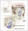
SLC26A4 genotypes and phenotypes associated with enlargement of the vestibular aqueduct
- PMID: 22116369
- PMCID: PMC3709178
- DOI: 10.1159/000335119
SLC26A4 genotypes and phenotypes associated with enlargement of the vestibular aqueduct
Abstract
Enlargement of the vestibular aqueduct (EVA) is the most common inner ear anomaly detected in ears of children with sensorineural hearing loss. Pendred syndrome (PS) is an autosomal recessive disorder characterized by bilateral sensorineural hearing loss with EVA and an iodine organification defect that can lead to thyroid goiter. Pendred syndrome is caused by mutations of the SLC26A4 gene. SLC26A4 mutations may also be identified in some patients with nonsyndromic EVA (NSEVA). The presence of two mutant alleles of SLC26A4 is correlated with bilateral EVA and Pendred syndrome, whereas unilateral EVA and NSEVA are correlated with one (M1) or zero (M0) mutant alleles of SLC26A4. Thyroid gland enlargement (goiter) appears to be primarily dependent on the presence of two mutant alleles of SLC26A4 in pediatric patients, but not in older patients. In M1 families, EVA may be associated with a second, undetected SLC26A4 mutation or epigenetic modifications. In M0 families, there is probably etiologic heterogeneity that includes causes other than, or in addition to, monogenic inheritance.
Copyright © 2011 S. Karger AG, Basel.
Figures


References
-
- Morton N. Genetic epidemiology of hearing impairment. Ann N Y Acad Sci. 1991;630:16–31. - PubMed
-
- Smith R, Bale JJ, White K. Sensorineural hearing loss in children. Lancet. 2005;365:879–890. - PubMed
-
- Gorlin RJ, Toriello H V, Cohen MM. Hereditary hearing loss and its syndromes. New York: Oxford University Press; 1995.
Publication types
MeSH terms
Substances
Grants and funding
LinkOut - more resources
Full Text Sources
Medical
