. 2012 Feb 1;93(2):291-301.
doi: 10.1093/cvr/cvr314. Epub 2011 Nov 23.

Pitx2 confers left morphological, molecular, and functional identity to the sinus venosus myocardium

Affiliations

Pitx2 confers left morphological, molecular, and functional identity to the sinus venosus myocardium

Grazia Ammirabile et al. Cardiovasc Res. .

Abstract

Aims: The sinus venous myocardium, comprising the sinoatrial node (SAN) and sinus horns (SH), is a region subject to congenital malformations and cardiac arrhythmias. It differentiates from symmetric bilateral mesenchymal precursors, but morphological, molecular, and functional left/right differences are progressively established through development. The role of the laterality gene Pitx2 in this process is unknown. We aimed to elucidate the molecular events driving left/right patterning in the sinus venosus (SV) myocardium by using a myocardial Pitx2 knockout mouse.

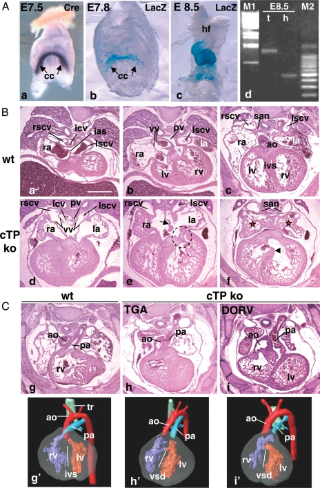
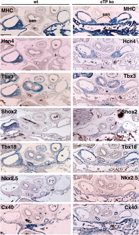
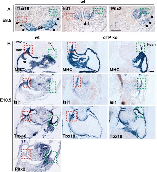
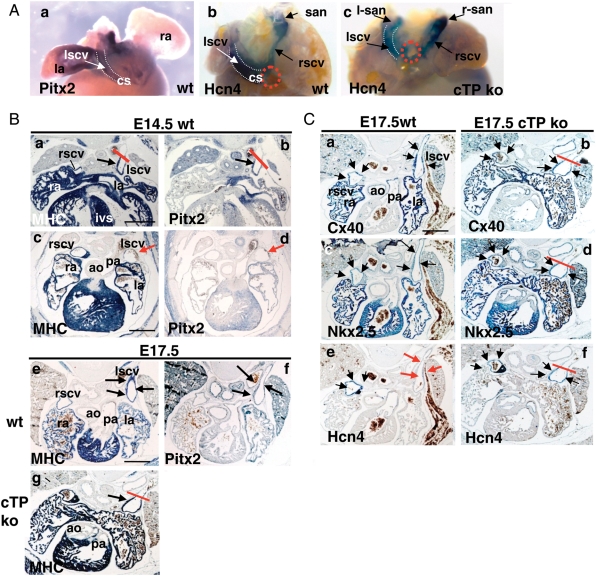
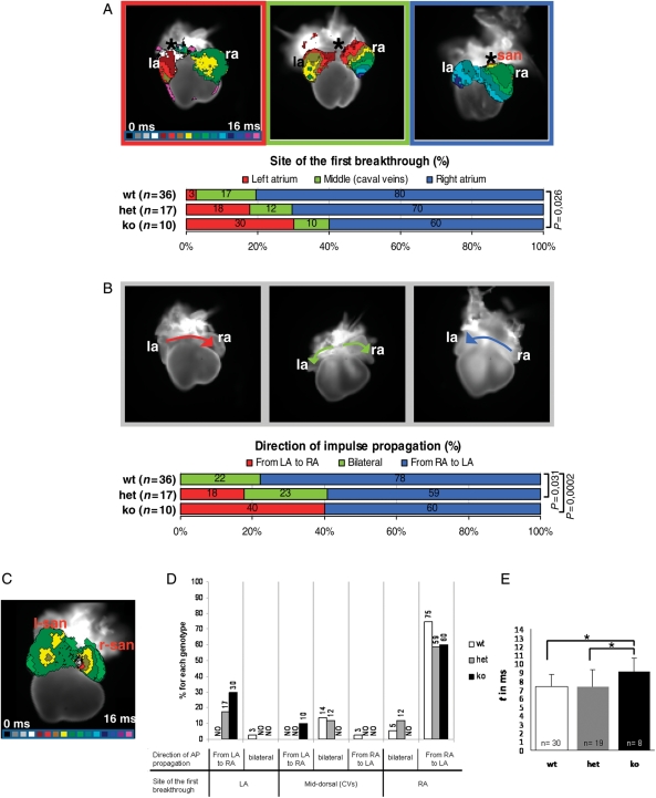
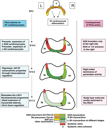
Methods and results: We generated a myocardial specific Pitx2 knockout model (cTP mice). cTP embryos present several features of Pitx2 null, including right atrial isomerism with bilateral SANs and symmetric atrial entrance of the systemic veins. By in situ hybridization and optical mapping analysis, we compared throughout development the molecular and functional properties of the SV myocardium in wt and mutant embryos. We observed that Pitx2 prevents the expansion of the left-SAN primordium at the onset of its differentiation into myocardium; Pitx2 promotes expansion of the left SH through development; Pitx2 dose-dependently represses the autorhythmic properties of the left SV myocardium at mid-gestation (E14.5); Pitx2 modulates late foetal gene expression at the left SH-derived superior caval vein.

Conclusion: Pitx2 drives left/right patterning of the SV myocardium through multiple developmental steps. Overall, Pitx2 plays a crucial functional role by negatively modulating a nodal-type programme in the left SV myocardium.


Figures

Figure 1
Figure 1
cTP mouse line characterization. (A) TnT Cre line characterization. Onset of Cre mRNA expression visualized by whole-mount ISH (Cre) (a) and onset of Cre activity (b), assessed by crossing TnT Cre with R26R mice (LacZ); (c) by E8.5 Cre activity is visible in the entire heart; (d) Pitx2 PCR on genomic DNA from trunk (t) and isolated heart (h) of a E8.5 cTP ko embryo to assess cardiac-specific recombination at the Pitx2 locus. M1: Lambda phage DNA, BstEII digested; M2: 100 bp ladder. From the embryonic trunk only the floxed allele is amplified (1232 bp band). In the corresponding heart only the 500 bp band is visible, indicating complete Cre-driven recombination. (B and C) H/E staining and MRI analysis of E14.5 wt and cTP ko hearts. Note that the sinoatrial region of the ko hearts presents the morphological features of RAI. Red stars in (f) show symmetrical pectinate muscles arrangement in atria. (e) Arrow indicates ASD, dotted circle indicates abnormal shape of the left atrioventricular valve; (f) arrowhead indicates VSD. (C) (h and i) Ventriculo-arterial alignment defects in cTP ko and (h′ and i′) their corresponding 3D MRI reconstructions. Cc, cardiac crescent; hf, headfolds; ias, interatrial septum; la, ra, left and right atrium; ivs, interventricular septum; san, sinoatrial node; lscv, rscv, left and right superior caval veins; vv, venous valves; icv, inferior caval vein; pv, pulmonary veins; lv, rv, left and right ventricle; ao, aorta; pa, pulmonary artery; tr, trachea. Scale bar: 0.5 mm.
Figure 2
Figure 2
The L-SAN of the cTP ko presents correct molecular pattern. ISH analysis of E14.5 wt and cTP ko embryos to assess L-SAN molecular signature. Note SAN expression of MHC, Hcn4, Tbx3, Shox2, Tbx18, and the negative staining with Nkx2.5 and CX40 antisense probes. Scale bar: 0.2 mm.
Figure 3
Figure 3
L-SAN mesenchymal precursors differentiate and expand into the left ectopic SAN of the cTP ko. (A) ISH of E8.5 wt embryos shows Tbx18 expression in SV mesenchymal precursors (arrows) and proepicardial organ (pe); Isl1 is bilaterally expressed in the second heart field (shf) overlapping Tbx18 at the SAN progenitors (dotted squares: green, right; red, left). Pitx2 expression delineates the left SV domain (arrows), including L-SAN progenitors (green dotted square). (B) At E10.5, cTP ko embryos present bilateral MHC, Isl1- and Tbx18-positive regions at the borders between left and right cardinal veins (lcv, rcv) and atria, identifying the early differentiated bilateral SAN. Scale bar: 0.1 mm.
Figure 4
Figure 4
Pitx2 modulates the LSCV developmental programme. (A) Whole-mount ISH of E14.5 wt hearts (dorsal view) showing expression of Pitx2 in LA and LSCV (a) and of Hcn4 in the entire SV myocardium, including the SAN (b). Red dotted circles (b and c) indicate ICV entrance. Note the absence of coronary sinus (cs) and symmetric CVs arrangement in the cTP ko (c). (B) Correlation between Pitx2 expression and MHC extension in the LSCV. In E14.5 wt hearts, note Pitx2 and MHC co-expression in the LSCV in four-chamber view sections (a and b) but not more distally (c and d); red bar indicates the border of probe detection. In E17.5 wt, MHC and Pitx2 expression is visible more distally (e and f), but in the cTP ko distal MHC extension is impaired (red bar in g). (C) The atrialization programme is partially impaired in the cTP ko. E17.5 wt embryos bilaterally express Cx40 (a) and Nkx2.5 (c) in the CVs, while Hcn4 expression is strongly down-regulated only in the LSCV (red arrows, e). In the cTP ko, Cx40 and Nkx2.5 signals are still present (b and d) in the reduced LSCV myocardial domain. Note the absence of Hcn4 down-regulation in the LSCV (black arrows in f). Scale bar: 0.5 mm.
Figure 5
Figure 5
Pitx2 represses left pacemaker activity within the SV myocardium of E14.5 embryos. (A) Site of first breakthrough. Top: prototypical maps (dorsal view) showing the three main atrial activation patterns recorded in isolated E14.5 hearts by optical mapping. The earliest activated region is indicated with an asterisk. Isochronal lines, delimiting regions activated within the same time frame, are spaced at 1 ms intervals; colour progression visualizes the advancing activation wavefront. Below: diagram showing the distribution of the activation patterns in the three genotypes. n = classified samples. (B) Direction of impulse propagation. Top: three representative examples of observed impulse propagation patterns; arrows depict the direction of electrical activation spread. Below: diagram illustrating pattern distribution within the genotypes. P-values <0.05 are indicated. (C) The L-SAN is functional. Activation map of a cTP ko heart showing the almost synchronous activation of the two SANs. (D and E) Atrial conduction properties in E14.5 wt and cTP mutants. (D) Correlation between the site of first atrial breakthrough and direction of impulse propagation. Columns indicate the combined distribution of atrial activation and propagation patterns within genotypes. Data are expressed as percentages; NO, never observed. (E) Atrial activation times. Data are presented as averages ± standard deviations; *P < 0.05.
Figure 6
Figure 6
Proposed model for Pitx2 action in left SV cardiomyocytes; hearts are shown in the dorsal view. Left columns summarize the temporal actions of Pitx2 in the developing SV region; right columns summarize the resulting morphogenetic and functional effects; v, embryonic ventricle; la, ra, left and right atrium.

References

    1. Christoffels VM, Mommersteeg MT, Trowe MO, Prall OW, de Gier-de Vries C, Soufan AT, et al. Formation of the venous pole of the heart from an Nkx2-5-negative precursor population requires Tbx18. Circ Res. 2006;98:1555–1563. - PubMed
    1. Sizarov A, Anderson RH, Christoffels VM, Moorman AF. Three-dimensional and molecular analysis of the venous pole of the developing human heart. Circulation. 2010;122:798–807. - PubMed
    1. Attenhofer Jost CH, Connolly HM, Danielson GK, Bailey KR, Schaff HV, Shen WK, et al. Sinus venosus atrial septal defect: long-term postoperative outcome for 115 patients. Circulation. 2005;112:1953–1958. - PubMed
    1. Lin WS, Tai CT, Hsieh MH, Tsai CF, Lin YK, Tsao HM, et al. Catheter ablation of paroxysmal atrial fibrillation initiated by non-pulmonary vein ectopy. Circulation. 2003;107:3176–3183. - PubMed
    1. Katritsis DG, Giazitzoglou E, Korovesis S, Karvouni E, Anagnostopoulos CE, Camm AJ. Conduction patterns in the cardiac veins: electrophysiologic characteristics of the connections between left atrial and coronary sinus musculature. J Interv Card Electrophysiol. 2004;10:51–58. - PubMed

Publication types