. 2011 Nov 23;3(110):110ra119.
doi: 10.1126/scitranslmed.3002823.

Rapid cancer detection by topically spraying a γ-glutamyltranspeptidase-activated fluorescent probe

Affiliations

Rapid cancer detection by topically spraying a γ-glutamyltranspeptidase-activated fluorescent probe

Yasuteru Urano et al. Sci Transl Med. .

Abstract

The ability of the unaided human eye to detect small cancer foci or accurate borders between cancer and normal tissue during surgery or endoscopy is limited. Fluorescent probes are useful for enhancing visualization of small tumors but are typically limited by either high background signal or the requirement for administration hours to days before use. We synthesized a rapidly activatable, cancer-selective fluorescence imaging probe, γ-glutamyl hydroxymethyl rhodamine green (gGlu-HMRG), with intramolecular spirocyclic caging for complete quenching. Activation occurs by rapid one-step cleavage of glutamate with γ-glutamyltranspeptidase (GGT), which is not expressed in normal tissue, but is overexpressed on the cell membrane of various cancer cells, thus leading to complete uncaging and dequenching of the fluorescence probe. In vitro activation of gGlu-HMRG was evident in 11 human ovarian cancer cell lines tested. In vivo in mouse models of disseminated human peritoneal ovarian cancer, activation of gGlu-HMRG occurred within 1 min of topically spraying the tumor, creating high signal contrast between the tumor and the background. The gGlu-HMRG probe is practical for clinical application during surgical or endoscopic procedures because of its rapid and strong activation upon contact with GGT on the surface of cancer cells.


Figures

Fig. 1.
Fig. 1.
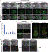
The gGlu-HMRG probe is specifically activated by GGT and retained in cancer cells. (A) Proposed mechanism of gGlu-HMRG activation and retention. gGlu-HMRG reacts with up-regulated GGT anchored in the plasma membrane of cancer cells. This interaction enzymatically cleaves the probe to yield a highly fluorescent and membrane-permeable product, HMRG, which accumulates in the lysosomes of cancer cells. (B) Aminopeptidase-sensitive fluorescence probes containing HMRG, such as gGlu-HMRG, fluoresce in their uncaged forms. (C) Changes in absorbance and fluorescence of gGlu-HMRG upon reacting with GGT. S/N, signal-to-noise ratio; a.u., arbitrary units. (D) Comparison of GGT and LAP activities in human cancer cells (SHIN3) and normal cells (HUVEC) in response to respective enzyme-specific probes gGlu-HMRG and Leu-HMRG. Scale bars, 25 μm. (E) Inhibitory effect of GGsTop in SHIN3 cells with gGlu-HMRG. Scale bar, 25 μm. (F) siRNA knockdown of GGT-1 in the SHIN3 cancer cells and activation of gGlu-HMRG. Data are mean fluorescence intensities (a.u.) ± SEM. (G) Stability of various aminopeptidase-activated probes and an esterase probe in 10% murine serum. The graph on the right is a magnified one of the graph on the left to show the difference in nonspecific activation of the aminopeptidase probes.
Fig. 2.
Fig. 2.
gGlu-HMRG probe illuminated SHIN3 tumors with minimal background in vivo. (A) Representative white light, fluorescence, and composite images of the mouse peritoneum (representative of three to five mice per group) 10 min after intraperitoneal injection of gGlu-HMRG, Leu-HMRG, FDA, Gly-HMRG, Ile-HMRG, or Phe-HMRG in a SHIN3 mouse model of disseminated ovarian cancer. (B) Tumor–to–normal tissue ratio of fluorescence intensity in the peritoneal cavity of SHIN3-disseminated mice with various activatable probes. (C) Ex vivo fluorescence imaging of the spread mouse mesentery of tumor-bearing mice and control mice with gGlu-HMRG. Scale bar, 1 cm. (D) A fluorescence image of the peritoneal cavity 10 min after intraperitoneal injection of gGlu-HMRG into the SHIN3 mouse model and a non–tumor-bearing normal mouse. Scale bars, 1 cm. (E) White light and fluorescence images of mouse peritoneal cavity and mesentery 10 min after intraperitoneal injection of gGlu-HMRG, with (+) or without (−) co-injection of GGsTop inhibitor. Scale bars, 1 cm.
Fig. 3.
Fig. 3.
gGlu-HMRG probe illuminates ovarian cancer cell lines in vitro. (A and B) Microscopic images of 11 human ovarian cancer cell lines 10 and 60 min after incubation (A) and flow cytometry results 60 min after incubation (B) with gGlu-HMRG. Scale bars, 50 μm.
Fig. 4.
Fig. 4.
gGlu-HMRG probe illuminates experimental tumors in vivo. (A) gGlu-HMRG fluorescence is detected endoscopically in implanted ovarian tumors in living mice over the course of 60 min (see videos S1 to S5 for ≤5 min after spraying gGlu-HMRG, and video S6 for 1 hour after spraying gGlu-HMRG). (B) Changes in tumor fluorescence signals in six ovarian cancer–bearing mice (n = 4 per group). Data are mean fluorescence intensities (a.u.) ± SEM of tumor nodule at different time points. Four of six peritoneal implants significantly increase in fluorescence signal over the first 10 min. *P < 0.01 compared to fluorescence signal immediately after spraying gGlu-HMRG, Wilcoxon matched-pairs test. (C) Within 90 s, gGlu-HMRG illuminates SHIN3 tumors growing in the mouse peritoneum (video S1). (D) Tiny ovarian cancer implants (~1 mm) were removed with forceps using fluorescence-guided endoscopy (video S8).
Fig. 5.
Fig. 5.
Spectral fluorescence images of six peritoneal ovarian cancers confirm in vivo endoscopic imaging results. In vivo fluorescence intensity 10 and 60 min after intraperitoneal gGlu-HMRG administration was evaluated with six peritoneal ovarian tumor models: SHIN3, SKOV3, OVCAR3, OVCAR4, OVCAR5, and OVCAR8. After establishing the intraperitoneal dissemination model, one mouse from each pair (four pairs per cell line) was injected intraperitoneally with gGlu-HMRG. Yellow arrowheads indicate tumor location. White arrows indicate pancreas location. Scale bar, 1 cm.
Fig. 6.
Fig. 6.
Sensitivity and specificity of gGlu-HMRG for detecting tumors about 1 mm in diameter. (A) No crosstalk of fluorescence signals between gGlu-HMRG and RFP using parent SHIN3 tumors (no RFP) and SHIN3-RFP tumors. (B and C) Fluorescent imaging 10 min after intraperitoneal (ip) injection of the gGlu-HMRG probe: microscopic (fluorescence microscope) (B) and macroscopic (whole-body fluorescence imager) (C). Scale bars, 1 mm (B) and 1 cm (C). (D) Scatter plot of the fluorescence intensities in each nodule. When RFP was used as a reference for location of SHIN3 cells (10 a.u.) and the threshold of green fluorescence signal was set at 4 a.u., the sensitivity and specificity of detecting SHIN3-RFP tumors with gGlu-HMRG were 100 and 100%, respectively, 10 min after injection.

Comment in

References

    1. Frangioni JV, New technologies for human cancer imaging. J. Clin. Oncol 26, 4012–4021 (2008). - PMC - PubMed
    1. Wallace MB, Kiesslich R, Advances in endoscopic imaging of colorectal neoplasia. Gastroenterology 138, 2140–2150 (2010). - PubMed
    1. Weissleder R, Pittet MJ, Imaging in the era of molecular oncology. Nature 452, 580–589 (2008). - PMC - PubMed
    1. Kobayashi H, Ogawa M, Alford R, Choyke PL, Urano Y, New strategies for fluorescent probe design in medical diagnostic imaging. Chem. Rev 110, 2620–2640 (2010). - PMC - PubMed
    1. Kobayashi H, Choyke PL, Target-cancer-cell-specific activatable fluorescence imaging probes: Rational design and in vivo applications. Acc. Chem. Res 44, 83–90 (2011). - PMC - PubMed

Publication types

LinkOut - more resources