. 2012 Jan 6;287(2):989-1006.
doi: 10.1074/jbc.M111.297150. Epub 2011 Nov 22.

Corneal antifibrotic switch identified in genetic and pharmacological deficiency of vimentin

Affiliations

Corneal antifibrotic switch identified in genetic and pharmacological deficiency of vimentin

Paola Bargagna-Mohan et al. J Biol Chem. .

Abstract

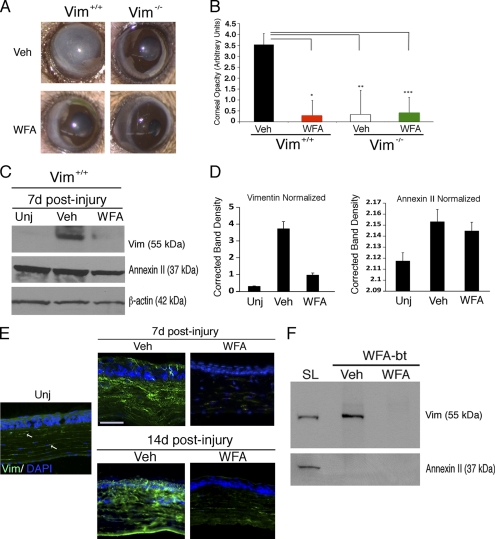
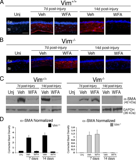
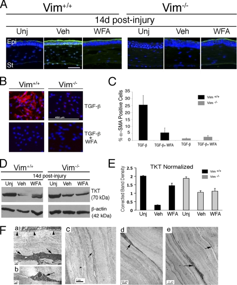
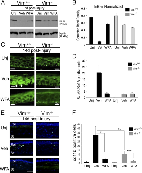
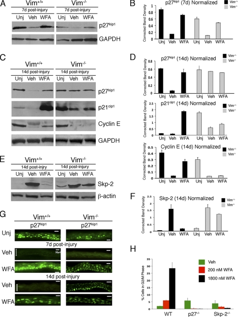
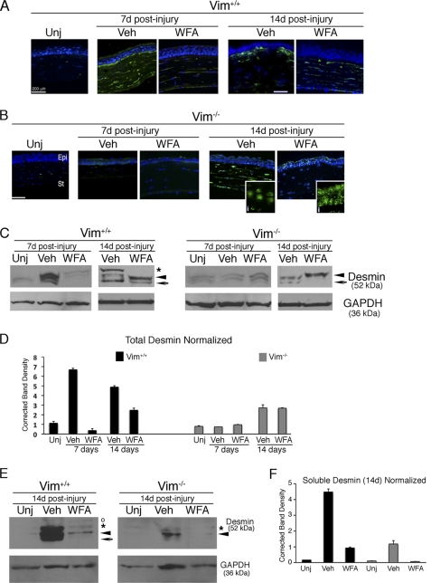
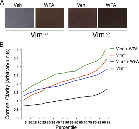
The type III intermediate filaments (IFs) are essential cytoskeletal elements of mechanosignal transduction and serve critical roles in tissue repair. Mice genetically deficient for the IF protein vimentin (Vim(-/-)) have impaired wound healing from deficits in myofibroblast development. We report a surprising finding made in Vim(-/-) mice that corneas are protected from fibrosis and instead promote regenerative healing after traumatic alkali injury. This reparative phenotype in Vim(-/-) corneas is strikingly recapitulated by the pharmacological agent withaferin A (WFA), a small molecule that binds to vimentin and down-regulates its injury-induced expression. Attenuation of corneal fibrosis by WFA is mediated by down-regulation of ubiquitin-conjugating E3 ligase Skp2 and up-regulation of cyclin-dependent kinase inhibitors p27(Kip1) and p21(Cip1). In cell culture models, WFA exerts G(2)/M cell cycle arrest in a p27(Kip1)- and Skp2-dependent manner. Finally, by developing a highly sensitive imaging method to measure corneal opacity, we identify a novel role for desmin overexpression in corneal haze. We demonstrate that desmin down-regulation by WFA via targeting the conserved WFA-ligand binding site shared among type III IFs promotes further improvement of corneal transparency without affecting cyclin-dependent kinase inhibitor levels in Vim(-/-) mice. This dissociates a direct role for desmin in corneal cell proliferation. Taken together, our findings illuminate a previously unappreciated pathogenic role for type III IF overexpression in corneal fibrotic conditions and also validate WFA as a powerful drug lead toward anti-fibrosis therapeutic development.


Figures

FIGURE 1.
FIGURE 1.
Control of vimentin expression favors corneal clarity in the alkaline burn injury model. Vim+/+ and Vim−/− mice were subjected to corneal chemical injury with limbal and corneal epithelial cell debridement and treated daily with DMSO (vehicle; Veh) or 2 mg/kg/day WFA by intraperitoneal injection for 7 and 14 days. A, representative images at d14 of Vim+/+ and Vim−/− whole eyes show dramatic reduction of corneal opacity in mice that have vimentin expression down-regulated either pharmacologically (WFA) or genetically (Vim−/−). B, quantification of corneal opacity by biomicroscopy at d14 using an opacity scale of 0 (clear) to 4 (opaque) (n = 8 samples/group). C, vimentin expression is down-regulated pharmacologically by WFA at d7 as shown by Western blot analysis of corneal buttons from uninjured (Unj), injured vehicle-treated (Veh), and WFA-treated Vim+/+ mice. Blots were probed sequentially with antibodies against vimentin (clone V9) and annexin II, and β-actin was used as loading control. Data are representative of two independent experiments (n = 4 mice/group). D, densitometric quantification of vimentin and annexin II normalized to β-actin using ImageJ software. E, immunofluorescence staining of vimentin expression (green) in thin tissue sections from corneas of uninjured, DMSO-treated, and WFA-treated Vim+/+ mice at d7 and d14. The image of the uninjured sample is enhanced compared with DMSO and WFA samples to reveal the low level of vimentin staining in corneal stromal keratocytes (white arrows). Nuclei are stained with DAPI (blue). Bars, 150 μm. Data are representative of two independent experiments (n = 4 mice/group). F, affinity isolation of WFA-bt-binding proteins from rabbit corneal fibroblasts. Corneal keratocytes were differentiated in vitro to wound fibroblasts and preincubated with DMSO (vehicle) or with 5 μm WFA for 30 min, and subsequently, both treatment groups were incubated with 5 μm WFA-bt for 2 h. Soluble protein lysates (SL) were obtained and subsequently purified over NeutrAvidin affinity columns and Western blotted by probing for vimentin and annexin II. A small amount of soluble lysate was included in parallel to demonstrate the presence of vimentin and annexin II in corneal cells. Error bars, S.D.
FIGURE 2.
FIGURE 2.
Corneal fibrosis is mediated by vimentin. Vim+/+ and Vim−/− mice were subjected to corneal alkali injury and treated daily with vehicle (Veh) or 2 mg/kg/day WFA by intraperitoneal injection for 7 and 14 days. A and B, immunofluorescence staining of α-SMA (red) in repairing corneas of Vim+/+ (A) and Vim−/− (B) mice treated with vehicle or WFA. Nuclei were stained with DAPI (blue). Epi, epithelium; St, stroma. Bar, 150 μm. Data are representative of two independent experiments (n = 8/group). C, immunoblot analysis of α-SMA expression in corneal tissues from uninjured and injured Vim+/+ and Vim−/− mice at d7 and d14 treated with vehicle or WFA; GAPDH was used as loading control. D, densitometric quantification of α-SMA expression normalized to GAPDH. Error bars, S.D.
FIGURE 3.
FIGURE 3.
WFA down-regulates TGF-β expression in injured corneas. A, Vim+/+ and Vim−/− mice were subjected to corneal alkali injury and treated daily with vehicle (Veh) or 2 mg/kg/day WFA by intraperitoneal injection for 14 days. Shown is immunofluorescence staining of TGF-β2 (green) in uninjured (Unj) and injured corneas of Vim+/+ and Vim−/− mice. Nuclei were stained with DAPI (blue). Data are representative of two independent experiments (n = 8/group). B, differentiation of Vim+/+ and Vim−/− fibroblasts to myofibroblasts with TGF-β treatment in the presence and absence of 500 nm WFA. Expression of α-SMA expression (red) was assessed by immunofluorescence staining and counterstained with DAPI to mark nuclei (blue). C, percentage of cells expressing α-SMA to total number of cell nuclei from three replicates. Data are representative of two independent experiments (n = 9/group). D, Western blot analysis of corneal tissues from uninjured and injured d14 Vim+/+ and Vim−/− mice treated with vehicle or WFA. Blots were probed with polyclonal antibody to TKT followed by β-actin. E, densitometric quantification of TKT normalized to β-actin using ImageJ. F, transmission electron microscopy of mouse corneas. Vim+/+ and Vim−/− mice were subjected to corneal alkali injury and treated daily with vehicle or 2 mg/kg/day WFA for 14 days. Transmission electron microscopy images of corneas from vehicle-treated Vim+/+ mice reveal well differentiated myofibroblasts having an abundance of rough endoplasmic reticuli (a, arrow). The epithelial basement membrane appears to be intact (a, arrowheads). The presence of PMNs is also observed in vehicle samples (b, arrow). In comparison, corneas of uninjured Vim+/+ mice reveal a typical staining pattern for keratocytes (c, arrow). Corneas of Vim+/+ injured mice treated with WFA reveal mostly keratocytes and wound fibroblasts (d, arrow), and myofibroblasts (not shown) were only rarely found. Injured Vim−/− corneas also contain keratocytes/fibroblasts, which were similarly observed in injured corneas of Vim−/− mice treated with WFA (d and e, arrows). Error bars, S.D.
FIGURE 4.
FIGURE 4.
WFA down-regulates expression of inflammatory markers during corneal repair. Vim+/+ and Vim−/− mice were subjected to corneal alkali injury and treated daily with vehicle (Veh) or 2 mg/kg/day WFA for 7 and 14 days. A, immunoblot analysis of corneal tissues from uninjured and injured Vim+/+ and Vim−/− mice at d7 treated with vehicle or WFA. B, densitometric quantification of IκB-α normalized to β-actin. C, immunolocalization of p65 RelA/NF-κB staining (green) in the epithelium of Vim+/+ and Vim−/− corneas at d14 showing nuclear localization of p65 (white arrows in the basal layer) in vehicle-treated Vim+/+ sample. Bar, 10 μm. D, percentage of cells showing p65/RelA staining in nuclei of epithelial cells was determined by comparing with DAPI staining (not shown). Data are representative of two independent experiments (n = 8/group). E, immunofluorescence staining of CD11b (green) in Vim+/+ and Vim−/− corneas at d14. Nuclei were counterstained with DAPI (blue). Epi, epithelium; St, stroma. Bar, 150 μm. Data are representative of two independent experiments (n = 8/group). F, numbers of CD11b+ cells detected in corneal tissue sections (n = 8/group). *, p = 0.0025; **, p = 0.010; ***, p = 0.0053. Error bars, S.D.
FIGURE 5.
FIGURE 5.
WFA cell cycle activity during corneal repair is mediated by vimentin. Vim+/+ and Vim−/− mice were subjected to corneal alkali injury and treated daily with vehicle (Veh) or 2 mg/kg/day WFA for 7 and 14 days. Corneal tissues were isolated, and equal amounts of protein extracts were subjected to Western blotting and probed sequentially with antibodies against p27Kip1, p21Cip1, and cyclin E (A and C). Blots from d14 samples were also probed with antibody to Skp2. E, densitometric quantification of proteins to GAPDH (B and D) and β-actin (F) was performed using ImageJ (National Institutes of Health). G, immunolocalization of p27Kip1 staining (green) in the epithelium of Vim+/+ and Vim−/− corneas at d7 and d14. Double arrowheads delimit the epithelium in injured (Veh) samples. Data are representative of two independent experiments (n = 8/group). Bar, 50 μm. H, WFA induces G2/M cell cycle arrest. Embryonic fibroblasts from wild-type (WT), p27Kip1-deficient, and Skp2-deficient mice were stimulated to proliferate in the presence of vehicle or WFA and subjected to flow cytometry for cell cycle analysis. Data are representative of two independent experiments. Error bars, S.D.
FIGURE 6.
FIGURE 6.
Molecular model of WFA-desmin. Atomic model of tetramers formed by head-to-tail dimers in the A11 and A22 configurations (A) that was assembled using a part of a graphic representation previously published (48). B, ribbon representation of the molecular dynamics-simulated desmin-WFA complex structure overlapped with vimentin-WFA complex. a, superposition of the desmin-WFA complex with the previously simulated vimentin-WFA complex (16) in which the ribbon structures of the desmin and vimentin are in white and blue, respectively. Amino acids that hydrogen-bond with ring A of the ligand are represented by their stick structures. b, ribbon structure of desmin-WFA showing hydrogen bonds between Gln-329 (Q329) and C1-ketone of WFA and between Asp-336 (D336) and the C4-hydroxyl group of WFA. The β-oriented 5,6-epoxide of WFA is positioned for nucleophilic attack by Cys-333 (C333) (yellow dotted arrow).
FIGURE 7.
FIGURE 7.
WFA down-regulates injury-induced desmin expression in healing corneas. Vim+/+ and Vim−/− mice were subjected to corneal alkali injury and treated daily with vehicle (Veh) or 2 mg/kg/day WFA for 7 and 14 days. A and B, temporal induction and localization of desmin (green) in repairing tissues of corneas from Vim+/+ (A) and Vim−/− (B) corneas from uninjured (Unj) and vehicle- and WFA-treated mice. Higher magnified images of Vim−/− corneal sections at d14 reveal a novel dotlike staining pattern for desmin in the epithelium (i) that appears nucleus-associated. Nuclei were stained with DAPI (blue). Epi, epithelium; St, stroma. Bar, 200 μm. Data are representative of two independent experiments (n = 8/group). C, total corneal tissue lysates were also prepared from d7 and d14 mice, subjected to Western blotting, and probed with desmin antibody. *, major 52-kDa protein species; arrowhead and arrow, lower molecular weight desmin species/variants differentially regulated. D, densitometric quantification of desmin in Vim+/+ and Vim−/− samples normalized to GAPDH. E, Western blot analysis of soluble desmin from corneas of d14 Vim+/+ and Vim−/− mice. *, major 52 kDa band; arrowhead and arrow, smaller sized desmin variants; open circle, a nonspecific band. F, densitometric quantification of soluble desmin in Vim+/+ and Vim−/− samples normalized to GAPDH. Error bars, S.D.
FIGURE 8.
FIGURE 8.
Computer-aided imaging analysis of corneal transparency. A, representative images of injured (vehicle; Veh) and WFA-treated corneas of Vim+/+ and Vim−/− mice showing opacity that ranked at the 50th percentile for corneal clarity. B, corneal clarity values for Vim+/+ and Vim−/− mice treated with and without WFA (n > 150 images/group) were plotted as a function of their percentile rank distributions to reveal the trends in healing for each group. Maximal clarity scores are at 4.0.

References

    1. Whitcher J. P., Srinivasan M., Upadhyay M. P. (2001) Corneal blindness. A global perspective. Bull. World Health Organ. 79, 214–221 - PMC - PubMed
    1. Salomao M. Q., Wilson S. E. (2009) Corneal molecular and cellular biology update for the refractive surgeon. J. Refract. Surg. 25, 459–466 - PMC - PubMed
    1. Taneri S., Weisberg M., Azar D. T. (2011) Surface ablation techniques. J. Cataract Refract. Surg. 37, 392–408 - PubMed
    1. Qazi Y., Wong G., Monson B., Stringham J., Ambati B. K. (2010) Corneal transparency: genesis, maintenance and dysfunction. Brain Res. Bull. 81, 198–210 - PMC - PubMed
    1. Schaffeld M., Herrmann H., Schultess J., Markl J. (2001) Vimentin and desmin of a cartilaginous fish, the shark Scyliorhinus stellaris. Sequence, expression patterns and in vitro assembly. Eur. J. Cell Biol. 80, 692–702 - PubMed

Publication types

MeSH terms