

Quantitative comparison of myocardial fiber structure between mice, rabbit, and sheep using diffusion tensor cardiovascular magnetic resonance
- PMID: 22117695
- PMCID: PMC3235060
- DOI: 10.1186/1532-429X-13-74
Quantitative comparison of myocardial fiber structure between mice, rabbit, and sheep using diffusion tensor cardiovascular magnetic resonance
Abstract
Background: Accurate interpretations of cardiac functions require precise structural models of the myocardium, but the latter is not available always and for all species. Although scaling or substitution of myocardial fiber information from alternate species has been used in cardiac functional modeling, the validity of such practice has not been tested.
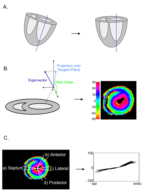
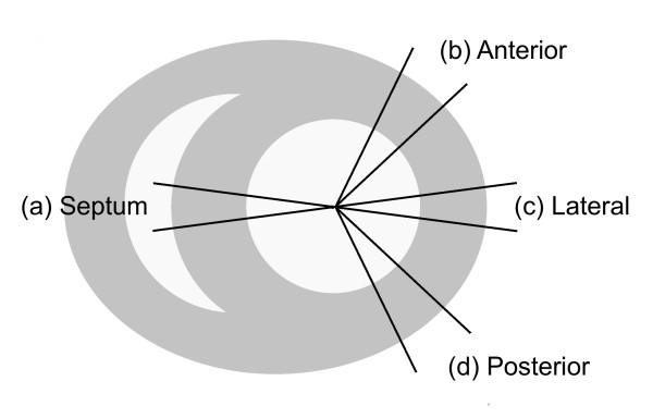
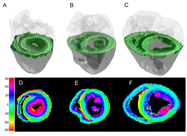
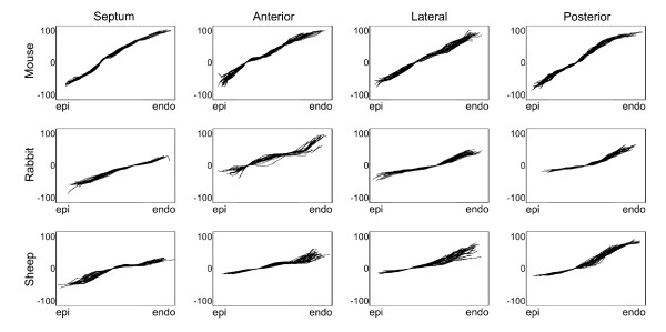
Methods: Fixed mouse (n = 10), rabbit (n = 6), and sheep (n = 5) hearts underwent diffusion tensor imaging (DTI). The myocardial structures in terms of the left ventricular fiber orientation helix angle index were quantitatively compared between the mouse rabbit and sheep hearts.
Results: The results show that significant fiber structural differences exist between any two of the three species. Specifically, the subepicardial fiber orientation, and the transmural range and linearity of fiber helix angles are significantly different between the mouse and either rabbit or sheep. Additionally, a significant difference was found between the transmural helix angle range between the rabbit and sheep. Across different circumferential regions of the heart, the fiber orientation was not found to be significantly different.
Conclusions: The current study indicates that myocardial structural differences exist between different size hearts. An immediate implication of the present findings for myocardial structural or functional modeling studies is that caution must be exercised when extrapolating myocardial structures from one species to another.
Figures
References
-
- Tranquillo JV, Hlavacek J, Henriquez CS. An integrative model of mouse cardiac electrophysiology from cell to torso. Europace. 2005;7(Suppl 2):56–70. - PubMed
-
- Niederer SA, Buist ML, Pullan AJ, Smith NP. Computing work in the ischemic heart. Conf Proc IEEE Eng Med Biol Soc. 2004;5:3646–3649. - PubMed
Publication types
MeSH terms
Grants and funding
LinkOut - more resources
Full Text Sources
