

Immortalized mesenchymal stem cells: an alternative to primary mesenchymal stem cells in neuronal differentiation and neuroregeneration associated studies
- PMID: 22118013
- PMCID: PMC3239243
- DOI: 10.1186/1423-0127-18-87
Immortalized mesenchymal stem cells: an alternative to primary mesenchymal stem cells in neuronal differentiation and neuroregeneration associated studies
Abstract
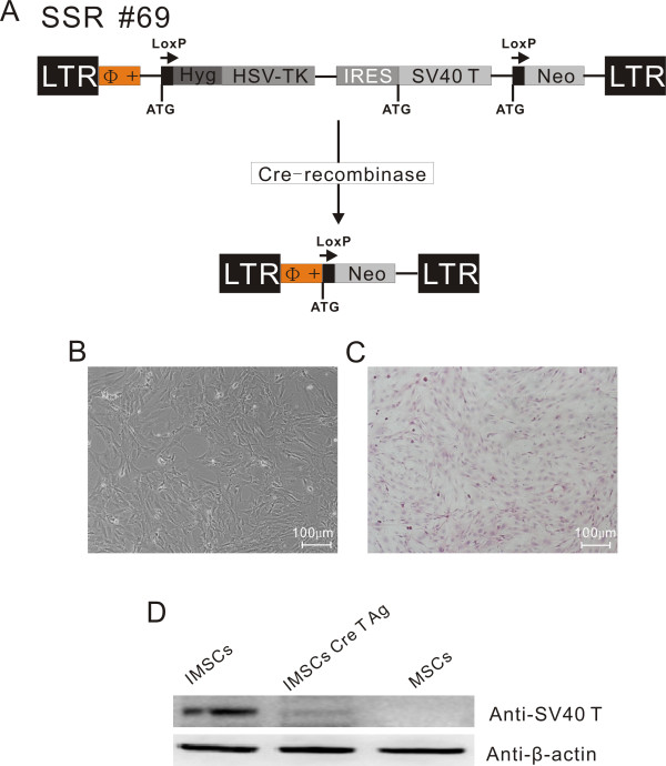
Background: Mesenchymal stem cells (MSCs) can be induced to differentiate into neuronal cells under appropriate cellular conditions and transplanted in brain injury and neurodegenerative diseases animal models for neuroregeneration studies. In contrast to the embryonic stem cells (ESCs), MSCs are easily subject to aging and senescence because of their finite ability of self-renewal. MSCs senescence seriously affected theirs application prospects as a promising tool for cell-based regenerative medicine and tissue engineering. In the present study, we established a reversible immortalized mesenchymal stem cells (IMSCs) line by using SSR#69 retrovirus expressing simian virus 40 large T (SV40T) antigen as an alternative to primary MSCs.
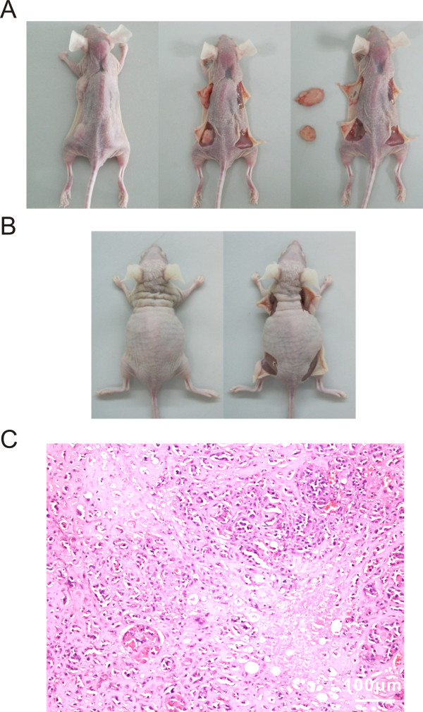
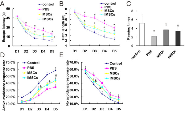
Methods: The retroviral vector SSR#69 expressing simian virus 40 large T (SV40T) antigen was used to construct IMSCs. IMSCs were identified by flow cytometry to detect cell surface makers. To investigate proliferation and differentiation potential of IMSCs, cell growth curve determination and mesodermal trilineage differentiation tests were performed. Neuronal differentiation characteristics of IMSCs were detected in vitro. Before IMSCs transplantation, we excluded its tumorigenicity in nude mice firstly. The Morris water maze tests and shuttle box tests were performed five weeks after HIBD models received cells transplantation therapy.
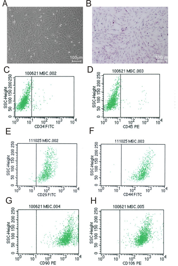
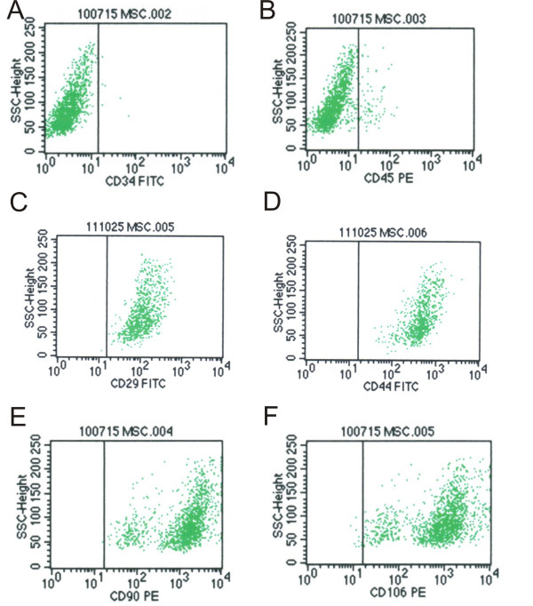
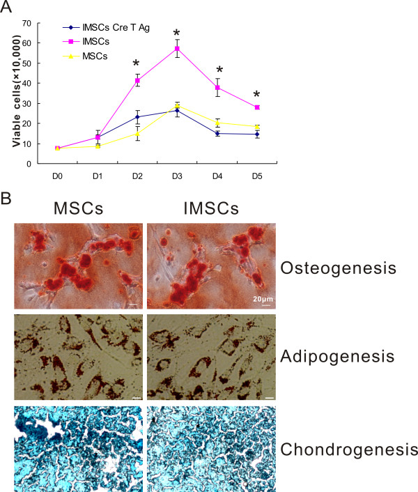
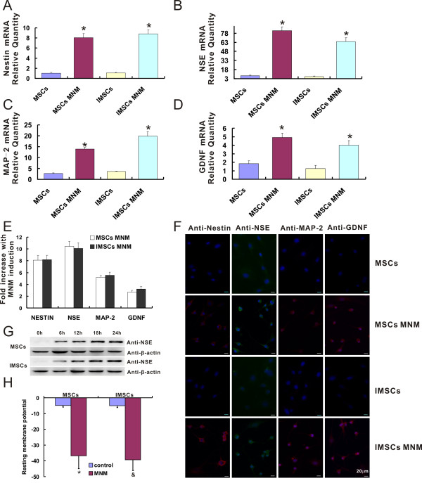
Results: In this study, reversible IMSCs were constructed successfully and had the similar morphology and cell surface makers as primary MSCs. IMSCs possessed better ability of proliferation and anti-senescence compared with primary MSCs, while maintained multilineage differentiation capacity. Neural-like cells derived from IMSCs had similar expressions of neural-specific genes, protein expression patterns and resting membrane potential (RMP) compared with their counterparts derived from primary MSCs. There was no bump formation in nude mice subcutaneously injected with IMSCs. IMSCs played same role as primary MSCs to improve learning ability and spatial memory of HIBD rats.
Conclusions: IMSCs not only retain their features of primary MSCs but also possess the ability of high proliferation and anti-senescence. IMSCs can definitely be induced to differentiate into neuronal cells in vitro and take the place of primary MSCs for cell transplantation therapy without tumorigenesis in vivo. The stable cell line is particularly useful and valuable as an alternative to MSCs in neuronal differentiation and neuroregeneration associated studies.
Figures
Similar articles
-
Human iPSC-derived MSCs (iMSCs) from aged individuals acquire a rejuvenation signature.Stem Cell Res Ther. 2019 Mar 18;10(1):100. doi: 10.1186/s13287-019-1209-x. Stem Cell Res Ther. 2019. PMID: 30885246 Free PMC article.
-
Reversibly immortalized human umbilical cord-derived mesenchymal stem cells (UC-MSCs) are responsive to BMP9-induced osteogenic and adipogenic differentiation.J Cell Biochem. 2018 Nov;119(11):8872-8886. doi: 10.1002/jcb.27140. Epub 2018 Aug 4. J Cell Biochem. 2018. PMID: 30076626 Free PMC article.
-
Generation of Mesenchymal Cell Lines Derived from Aged Donors.Int J Mol Sci. 2021 Oct 1;22(19):10667. doi: 10.3390/ijms221910667. Int J Mol Sci. 2021. PMID: 34639008 Free PMC article.
-
hiPSC-derived iMSCs: NextGen MSCs as an advanced therapeutically active cell resource for regenerative medicine.J Cell Mol Med. 2016 Aug;20(8):1571-88. doi: 10.1111/jcmm.12839. Epub 2016 Apr 21. J Cell Mol Med. 2016. PMID: 27097531 Free PMC article. Review.
-
Methods to produce induced pluripotent stem cell-derived mesenchymal stem cells: Mesenchymal stem cells from induced pluripotent stem cells.World J Stem Cells. 2021 Aug 26;13(8):1094-1111. doi: 10.4252/wjsc.v13.i8.1094. World J Stem Cells. 2021. PMID: 34567428 Free PMC article. Review.
Cited by
-
Neural Tissue-Like, not Supraphysiological, Electrical Conductivity Stimulates Neuronal Lineage Specification through Calcium Signaling and Epigenetic Modification.Adv Sci (Weinh). 2024 Sep;11(35):e2400586. doi: 10.1002/advs.202400586. Epub 2024 Jul 10. Adv Sci (Weinh). 2024. PMID: 38984490 Free PMC article.
-
Adenovirus-mediated RAR-β over-expression enhances ATRA-induced neuronal differentiation of rat mesenchymal stem cells.Arch Med Sci. 2013 Apr 20;9(2):314-22. doi: 10.5114/aoms.2012.31410. Epub 2012 Nov 6. Arch Med Sci. 2013. PMID: 23671444 Free PMC article.
-
A prospect of cell immortalization combined with matrix microenvironmental optimization strategy for tissue engineering and regeneration.Cell Biosci. 2019 Jan 5;9:7. doi: 10.1186/s13578-018-0264-9. eCollection 2019. Cell Biosci. 2019. PMID: 30627420 Free PMC article. Review.
-
Comparative capability of menstrual blood versus bone marrow derived stem cells in neural differentiation.Mol Biol Rep. 2017 Feb;44(1):169-182. doi: 10.1007/s11033-016-4095-7. Epub 2016 Dec 15. Mol Biol Rep. 2017. PMID: 27981446
-
Efficient Transformation of Primary Human Mesenchymal Stromal Cells by Adenovirus Early Region 1 Oncogenes.J Virol. 2016 Dec 16;91(1):e01782-16. doi: 10.1128/JVI.01782-16. Print 2017 Jan 1. J Virol. 2016. PMID: 27795433 Free PMC article.
References
-
- Jiang Y, Jahagirdar BN, Reinhardt RL, Schwartz RE, Keene CD, Ortiz-Gonzalez XR, Reyes M, Lenvik T, Lund T, Blackstad M, Du J, Aldrich S, Lisberg A, Low WC, Largaespada DA, Verfaillie CM. Pluripotency of mesenchymal stem cells derived from adult marrow. Nature. 2002;418(6893):41–9. doi: 10.1038/nature00870. - DOI - PubMed
-
- Mezey E, Chandross KJ, Harta G, Maki RA, McKercher SR. Turning blood into brain: cells bearing neuronal antigens generated in vivo from bone marrow. Science. 2000;290(5497):1779–82. - PubMed
-
- Ishii K, Yoshida Y, Akechi Y, Sakabe T, Nishio R, Ikeda R, Terabayashi K, Matsumi Y, Gonda K, Okamoto H, Takubo K, Tajima F, Tsuchiya H, Hoshikawa Y, Kurimasa A, Umezawa A, Shiota G. Hepatic differentiation of human bone marrow-derived mesenchymal stem cells by tetracycline-regulated hepatocyte nuclear factor 3beta. Hepatology. 2008;48(2):597–606. doi: 10.1002/hep.22362. - DOI - PubMed
Publication types
MeSH terms
LinkOut - more resources
Full Text Sources
Other Literature Sources
