. 2011 Nov 23;18(11):1369-78.
doi: 10.1016/j.chembiol.2011.08.019.

Arginylation and methylation double up to regulate nuclear proteins and nuclear architecture in vivo

Affiliations

Arginylation and methylation double up to regulate nuclear proteins and nuclear architecture in vivo

Sougata Saha et al. Chem Biol. .

Abstract

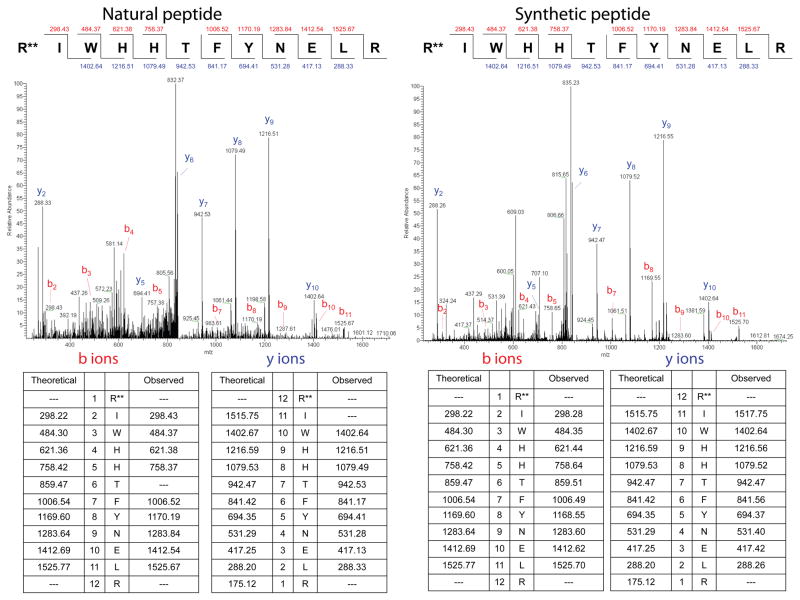
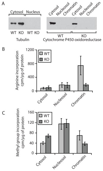
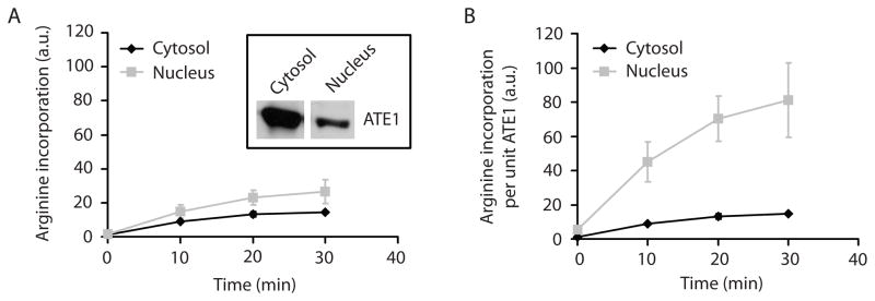
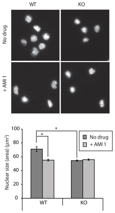
Protein arginylation and arginine methylation are two posttranslational modifications of emerging importance that involve Arg residues and their modifications. To test a hypothesis that posttranslationally added arginines can be methylated, we used high-precision mass spectrometry and metabolic labeling to find whether posttranslationally added arginines can serve as methylation sites. We identified a number of proteins in vivo, on which posttranslationally added Arg have undergone mono- and dimethylation. This double modification predominantly affects the chromatin-containing nuclear fraction and likely plays an important regulatory role in chromatin-associated proteins. Moreover, inhibition of arginylation and Arg methylation results in a significant reduction of the nucleus size in cultured cells, suggesting changes in chromatin compaction and nuclear architecture. Our findings suggest a functional link between protein regulation by arginylation and methylation that affects nuclear structure in vivo.


Figures

Figure 1
Figure 1. MS/MS spectra of the experimentally identified dimethyl-arginylated actin peptide (NP-033736.1) (left, natural peptide) and synthetic standard peptide containing N-terminal dimethylated Arg (right, dimethyl groups in both are denoted with double stars)
Peptide sequences and b and y ion masses are shown on the top right for each spectrum and in the tables underneath. See Supplemental Table S1 for the search parameters. See also Figure S1.
Figure 2
Figure 2. Arg and methyl group incorporation into different subcellular fractions
A. Western blot of the cytosolic and nuclear fractions with the cytoplasmic marker tubulin (left) and the ER marker cytochrome P450 oxidoreductase indicate that purified nuclear fractions are devoid of cytosolic and ER contaminants. All the fractions for Western blotting were normalized by total protein amount. B. In vivo incorporation of 3H-Arg into cycloheximide and chloramphenicol -treated wild type (WT) and Ate1 knockout (KO) cells presented as c.p.m. per μg of protein. Error bars +/− SEM, n = 4. Chromatin fraction p value is 0.0874. C. In vivo incorporation of 3H-Met (the source of the methyl groups) into cycloheximide/chloramphenicol-treated WT and Ate1 KO cells presented as c.p.m. per μg of protein. Error bars +/− SEM, n = 3. Cytosol and chromatin fraction p values are 0.016 and 0.1, respectively. See also Figures S2 and S3.
Figure 3
Figure 3. ATE1 is less abundant but more active in the nucleus
A. Inset: Western blot showing ATE1 levels in the cytosol and nucleus in the samples loaded at the equal protein level. Chart: 3H-Arg incorporation by endogenous ATE1 into cellular proteins in cytosolic and nuclear extracts at different time points. Error bars +/− SEM, n = 2. B. The same curves as in A normalized to the ATE1 levels detected by the Western blots shown in A, inset. ATE1 activity per molecule is significantly higher in the nucleus than in the cytosol. See also Figure S4.
Figure 4
Figure 4. Structures of some of the arginylated and arginylated/methylated proteins identified in this study
The position of the postranslationally added Arg or methyl-Arg residues in the represented structures are modeled by incorporating the Arg or methyl-Arg in the primary structure of the protein. Postranslationally added arginines are marked with green and the side chain amino groups are in blue. The methyl groups in arginine are marked by green spots attached to the blue amino group. Arginylation sites are shown with red arrowheads and arginylation/methylation sites – with blue arrows. A. Nucleosome (structures shown are selected from the complex between nucleosome core particle (H3, H4, H2a, H2b) and 146 Bp Long DNA fragment, PDB identifier: 1AOI). The position of individual histones (marked on top) and the entire nucleosome assembly (marked ‘Nucleosome in the top rightmost panel) are shown in the topmost panels. The modified sites are located on the DNA binding and histone interaction surfaces likely to affect nucleosome assembly. The distances between the nitrogen atoms of added Arg or methyl-Arg and the nearest oxygen atoms of phosphate backbone of the DNA molecule are represented in the inset images a, b, c, and d and marked by black dotted lines. B. Hsp90 dimer (structure shown is of S. cerevisiae Hsp90, PDB identifier: 2CG9). Modified sites are located on the protein surface and near subunit interaction sites. Inset images e and f show a detailed image of the modified regions. C. U5snRNP specific protein (structure shown is of a homologus S. cerevisiae protein in complex with rRNA obtained by selecting the relevant structures from the S. cerevisiae ribosomal 80s-Eef2-sordarin complex, PDB identifier: 1S1H). Inset image g shows a detailed image of the modified regions. See also Supplemental Figures S4 for additional structures. See also Figure S5, Supplemental Table 2 and Supplemental Datasets S1 and S2.
Fig. 5
Fig. 5. Arginylation methylation regulates nuclear size
A. DAPI stained untreated or AMI 1 treated WT and Ate1 KO MEF nucleus. Bar 25 μm. B. Quantification of nuclear size observed in the experiment presented in panel A from following number of nucleus analyzed. WT, n= 41; Ate1 KO, n=96; WT+ AMI 1, n= 84; Ate1 KO+ AMI 1, n=150. Error bar +/− SEM. t-test p value <0.0001 for WT vs Ate1 KO and WT+ AMI 1.

References

    1. Bedford MT, Richard S. Arginine Methylation: An Emerging Regulator of Protein Function. Molecular Cell. 2005;18:263–272. - PubMed
    1. Emsley P, Cowtan K. Coot: model-building tools for molecular graphics. Acta Crystallogr D Biol Crystallogr. 2004;60:2126–2132. - PubMed
    1. Fackelmayer FO. Protein arginine methyltransferases: guardians of the Arg? Trends Biochem Sci. 2005;30:666–671. - PubMed
    1. Grummt I. Actin and myosin as transcription factors. Curr Opin Genet Dev. 2006;16:191–196. - PubMed
    1. Kaji H. Amino-terminal arginylation of chromosomal proteins by arginyl-tRNA. Biochemistry. 1976;15:5121–5125. - PubMed

Publication types

LinkOut - more resources