. 2011 Nov 23;18(11):1442-52.
doi: 10.1016/j.chembiol.2011.08.011.

Titration-based screening for evaluation of natural product extracts: identification of an aspulvinone family of luciferase inhibitors

Affiliations

Titration-based screening for evaluation of natural product extracts: identification of an aspulvinone family of luciferase inhibitors

Patricia G Cruz et al. Chem Biol. .

Abstract

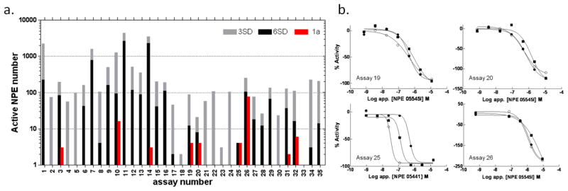
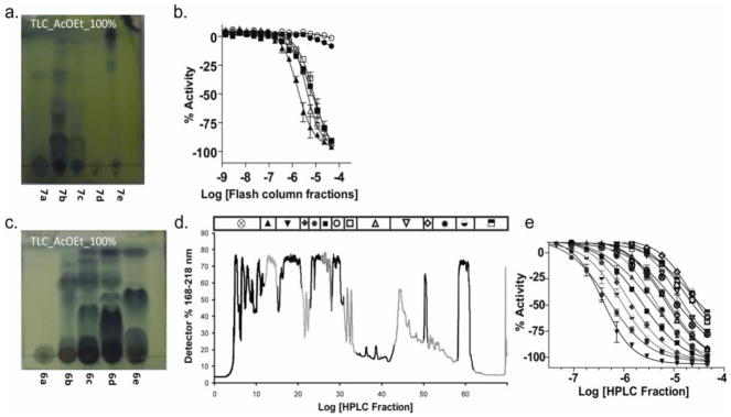
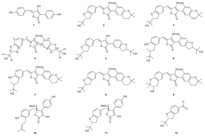
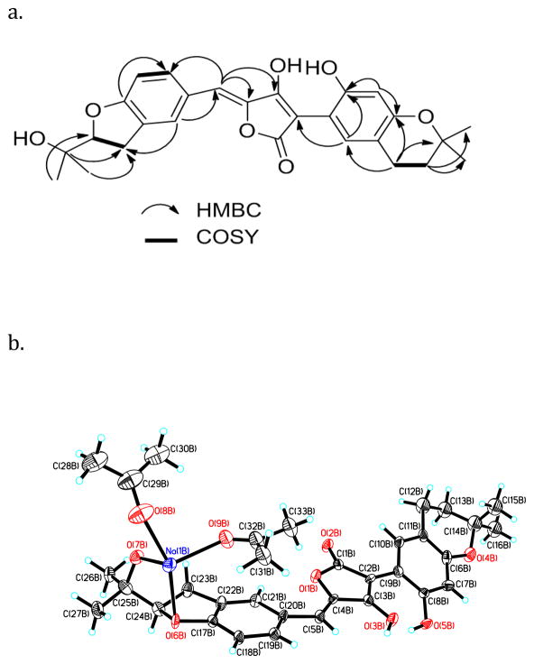
The chemical diversity of nature has tremendous potential for the discovery of molecular probes and medicinal agents. However, sensitivity of HTS assays to interfering components of crude extracts derived from plants, and macro- and microorganisms has curtailed their use in lead discovery. Here, we describe a process for leveraging the concentration-response curves obtained from quantitative HTS to improve the initial selection of "actives" from a library of partially fractionated natural product extracts derived from marine actinomycetes and fungi. By using pharmacological activity, the first-pass CRC paradigm improves the probability that labor-intensive subsequent steps of reculturing, extraction, and bioassay-guided isolation of active component(s) target the most promising strains and growth conditions. We illustrate how this process identified a family of fungal metabolites as potent inhibitors of firefly luciferase, subsequently resolved in molecular detail by X-ray crystallography.


Figures

Figure 1
Figure 1. Activity analysis of NPEs in various assays
a. Bar chart summarizing active NPEs in assays ordered by format (1–15 are fluorogenic; 16–18 are fluorescent polarization; 19–30 are bioluminescent; 31–32 ALPHA based chemluminescent; 33–34 use fluorescent protein expression and 35 is an absorbance output). The number of active NPEs per assay are indicated on the y-axis, where red represents a class 1a CRC (as previously defined [11]), while 3SD and 6SD cutoffs for activity at a single apparent concentration of 10 μM are given by the grey and black bars. b. Plots showing qHTS class 1a CRC results from assays selected for further analysis (see also Figure S2b). Assays 19, 20 and 26 displayed activity from a common strain, NPE 05545. Additional assay details can be found Table S1
Figure 2
Figure 2. Bioactivity guided de-replication of NPE 05545
a. Thin layer chromatography analysis of flash column fractions from an acetone extract of XAD-16-bound culture extract from strain 05545. b. Activity of flash column fractions shown in a in FLuc enzyme assay (open square, X+A; solid triangle, 7a; open triangle, 7b, closed square, 7c; solid circle, 7d; open circle, 7e). c. Thin layer chromatography analysis of flash column fractions from an independent culture of 05545. d. Reverse HPLC of the components of flash column fraction 6c in panel C. e. Activity from the HPLC separation shown in panel C.
Figure 3
Figure 3. Structures of isolated metabolites
Aspulvinone E (1), aspulvinone F (2), aspulvinone H (3), aspulvinone I-CR (4), aspulvinone J-CR (5), aspulvinone K-CR (6), aspulvinone L-CR (7), aspulvinone M-CR (8), aspulvinone N-CR (9), butyrolactone I (10), butyrolactone III (11) and benzofuran (12). 1H, 13C NMR, COSY and HSQC spectra for compounds 2, 49 are provided in Figures S4-S31 and Tables S3 and S4.
Figure 4
Figure 4. NMR and X-ray analysis of aspulvinone F (2)
a. Key 1H-1H COSY and HMBC correlations observed in 2, b. X-ray structure of 2. Perspective views showing 50% probability displacement ellipsoids of an independent aspulvinone F molecule. Na+ is chelated with four oxygens, two from acetone molecules and two from aspulvinone F (2). Carbon atoms are shown in black, oxygen atoms in red, protons in white, and Na+ in blue.
Figure 5
Figure 5. Structure of FLuc containing bound aspulvinone J-CR (5)
a. View of 5 (blue and red cylinders) in the active site of luciferase (grey ribbons). The ATP and luciferin binding regions are colored magenta and green respectively. b. Two views of the Fo-Fc electron density map for 5 contoured at 3sσ. c. Hydrogen bonding between 5 (grey/red) and luciferase (cyan). Water molecules are drawn as red spheres. Direct contacts between luciferase and 5 are shown as dashed lines and water mediated interactions are indicated by the solid lines. Crystallographic data for 5 can be found in Table S2. Coordinates and structure factors have been deposited to the Protein Databank with the accession code 3RIX.
Figure 6
Figure 6. Superposition of aspulvinone J-CR (5) and PTC124-AMP adduct within FLuc binding pocket
Protein is shown in ribbon representation and the binding pocket is depicted by molecular surface, 5 is shown in green and PTC124-AMP is shown in cyan. Phe247 is shown in grey. This figure was prepared with the program VIDA (OpenEye Scientific Software).

References

    1. Adams PD, et al. PHENIX: a comprehensive Python-based system for macromolecular structure solution. Acta crystallographica. 2010;66:213–221. - PMC - PubMed
    1. Auld DS, Lovell S, Thorne N, Lea WA, Maloney DJ, Shen M, Rai G, Battaile KP, Thomas CJ, Simeonov A, et al. Molecular basis for the high-affinity binding and stabilization of firefly luciferase by PTC124. Proc Natl Acad Sci U S A. 2010;107:4878–4883. - PMC - PubMed
    1. Auld DS, Southall NT, Jadhav A, Johnson RL, Diller DJ, Simeonov A, Austin CP, Inglese J. Characterization of chemical libraries for luciferase inhibitory activity. J Med Chem. 2008a;51:2372–2386. - PubMed
    1. Auld DS, Thorne N, Maguire WF, Inglese J. Mechanism of PTC124 activity in cell-based luciferase assays of nonsense codon suppression. Proc Natl Acad Sci U S A. 2009a;106:3585–3590. - PMC - PubMed
    1. Auld DS, Thorne N, Nguyen DT, Inglese J. A specific mechanism for nonspecific activation in reporter-gene assays. ACS Chem Biol. 2008b;3:463–470. - PMC - PubMed

Publication types

MeSH terms

Associated data

LinkOut - more resources