. 2012 Mar;12(2):294-304.
doi: 10.1016/j.mito.2011.11.002. Epub 2011 Nov 20.

Mice deleted for heart-type cytochrome c oxidase subunit 7a1 develop dilated cardiomyopathy

Affiliations

Mice deleted for heart-type cytochrome c oxidase subunit 7a1 develop dilated cardiomyopathy

Maik Hüttemann et al. Mitochondrion. 2012 Mar.

Abstract

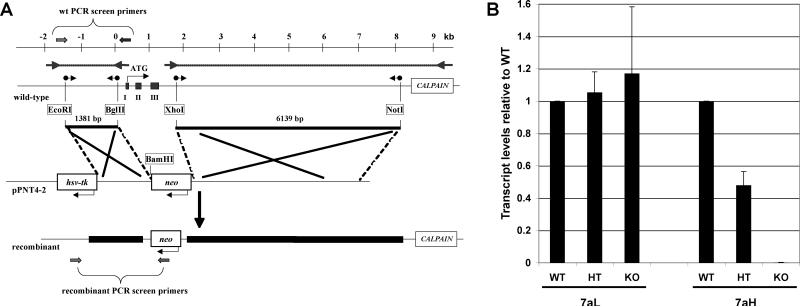
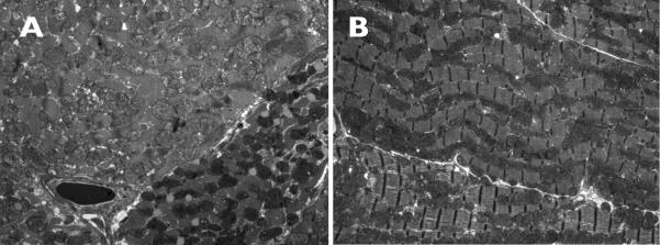
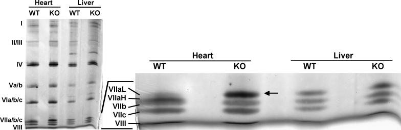
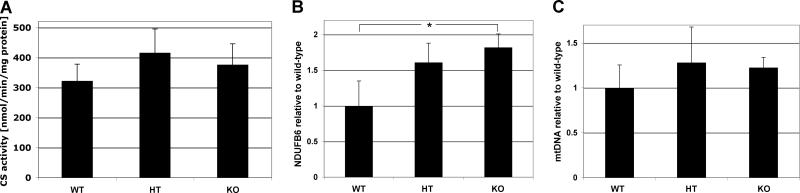
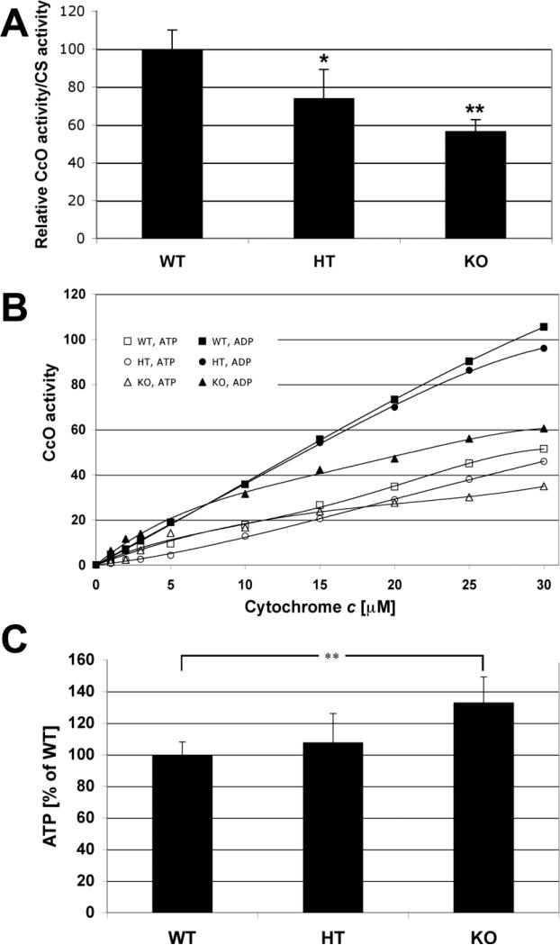
Subunit 7a of mouse cytochrome c oxidase (Cox) displays a contractile muscle-specific isoform, Cox7a1, that is the major cardiac form. To gain insight into the role of this isoform, we have produced a new knockout mouse line that lacks Cox7a1. We show that homozygous and heterozygous Cox7a1 knockout mice, although viable, have reduced Cox activity and develop a dilated cardiomyopathy at 6 weeks of age. Surprisingly, the cardiomyopathy improves and stabilizes by 6 months of age. Cox7a1 knockout mice incorporate more of the "liver-type" isoform Cox7a2 into the cardiac Cox holoenzyme and, also surprisingly, have higher tissue ATP levels.


Figures

Fig. 1
Fig. 1. Cox7a1 knockout mouse
A) Schematic representation of the knockout strategy. The PCR-based amplification of the 3′- and 5′-Cox7a1 regions (outer PCR: arrows with dotted lines) was followed by a nested PCR with primers containing the indicated restriction sites (sphere-tailed arrows). Two fragments are generated, the 800-bp promoter region to the end of intron I (1381-bp fragment), and the 6139-bp region spanning from intron III to the calpain gene. These fragments were cloned into the respective sites of the pPNT vector (pPNT7a1), leading to a replacement of exons I to III by the neo cassette in the recombinant. B) Relative transcript level of Cox7a isoforms in each genotype. Determination by qPCR is relative to Gapdh.
Fig 2
Fig 2. Electron microscope images of cardiac sections
Tissues from (A) Cox7a1+/+ and (B) Cox7a1-/- mouse hearts were examined in thin sections after staining with osmium tetroxide and uranylic acid as described in Methods. Darker mitochondria (white arrowhead) of unknown etiology were present in the -/- hearts in greater numbers than the more usual structures seen in wild-type sections (white arrow).
Fig. 3
Fig. 3. Lack of Cox7a1 (Cox7aH) in the Cox7a1 knockouts is complemented by increased Cox7a2 (Cox7aL) isoform incorporation into the Cox holoenzyme
For 2D-gel electrophoresis, heart and liver mitochondria were isolated from Cox7a1 knockout (KO) and wild-type mice (WT) from two pooled hearts and a liver each. OxPhos complexes were solubilized using dodecyl maltoside and run on BisTris 5-13% blue native PAGE in the first dimension. Bands corresponding to Cox were dissected and resolved by 6 M urea/18% SDS-PAGE (Lee et al., 2005), optimized for the separation of smaller Cox subunits 7 (VII) a, b, c, and 8 (VIII). Cox subunits were visualized by silver staining. In the absence of Cox7a1 in the knockouts, significantly increased protein levels of the liver-type isoform were observed (arrow).
Fig. 4
Fig. 4. Mitochondrial components trend upwards in heterozygote and null mice
A) Citrate synthase (CS) activity was measured as described in Materials and Methods. There was a trend in which CS activity was 29% and 17% increased in the heterozygotes and the Cox7a1 knockouts, respectively. However, differences were not statistically significant (p=0.13 WT-HT; p=0.27, WT-KO) (n=4). B) Ndufb6 gene expression was measured by qPCR as described. *, p<0.05. C) MtDNA content of Cox7a1+/+, Cox7a1+/- and Cox7a1-/- was determined by qPCR as described in Materials and Methods, relative to a single copy nuclear gene (mouse β-2 microglobulin) using total DNA.
Fig. 5
Fig. 5. Cox activity and ATP levels in Cox7a1 heterozygous and knockout mice
A) Cox activity is decreased in hearts of heterozygous and knockout mice. Cox activity of heart homogenates was determined with the spectrophotometric method and standardized to citrate synthase activity. Wild-type (WT) was set to 100%. Heterozygotes (HT) and Cox7a1 knockout mice had 26% and 53% reduced Cox activities, respectively. Note that measurements were performed in the absence of allosteric Cox activity modulators ADP and ATP (n=4; *, p<0.5; **, p<0.01). B) Cox activity is decreased in heart tissue of Cox7a1 knockout mice. Cox activity was determined in heart tissue homogenates of wild-type (WT, squares), heterozygote, (HT, circles), and Cox7a1 knockout mice (KO, triangles) in the presence of allosteric Cox inhibitor ATP (open symbols) and allosteric activator ADP (closed symbols) with the polarographic method by increasing the amount of substrate cytochrome c. Cox activity is defined as consumed O2 [nmol]/min/protein [mg]. Shown are representative measurements (n = 4 each; standard deviation < 5% at maximal turnover). C) ATP levels are increased in the Cox7a1 knockout mice. ATP concentrations of heart median cross-sections were determined with the bioluminescence method. ATP levels are 7% and 33% increased in the heterozygotes (HT) and Cox7a1 knockouts (KO) in comparison to the wild-types (WT), respectively. ATP at 100% is equivalent to 170 μg ATP/mg of solubilized protein (n = 6 animals in each group, measured in triplicates each; **, p < 0.01).
Fig 6
Fig 6. Echo analysis of wild-type and Cox7a1-/- mice
Two-dimensional and M-mode imaging of the left ventricle in the long-axis view demonstrates LV chamber enlargement and impaired contractility for Cox7a1-/- mice. Spectral Tissue Doppler imaging of the LV lateral wall demonstrates diminished e’ wave velocities and a’ wave for Cox7a1-/- mice, consistent with LV diastolic dysfunction.

References

    1. Acin-Perez R, Bayona-Bafaluy MP, Bueno M, Machicado C, Fernandez-Silva P, Perez-Martos A, Montoya J, Lopez-Perez MJ, Sancho J, Enriquez JA. An intragenic suppressor in the cytochrome c oxidase I gene of mouse mitochondrial DNA. Hum. Mol. Genet. 2003;12(3):329–339. - PubMed
    1. Arber S, Hunter JJ, Ross J, Jr., Hongo M, Sansig G, Borg J, Perriard J-C, Chien KR, Caroni P. MLP-deficient mice exhibit a disruption of cardiac cytoarchitectural organization, dilated cardiomyopathy, and heart failure. Cell. 1997;88(3):393–403. - PubMed
    1. Bonne G, Seibel P, Possekel S, Marsac C, Kadenbach B. Expression of human cytochrome-c oxidase subunits during fetal development. Eur. J. Biochem. 1993;217(3):1099–1107. - PubMed
    1. Burelle Y, Khairallah M, Ascah A, Allen BG, Deschepper CF, Petrof BJ, Des Rosiers C. Alterations in mitochondrial function as a harbinger of cardiomyopathy: Lessons from the dystrophic heart. J. Mol. Cell. Cardiol. 2010;48(2):310–321. - PMC - PubMed
    1. Casali C, Damati G, Bernucci P, Debiase L, Autore C, Santorelli FM, Coviello D, Gallo P. Maternally inherited cardiomyopathy: Clinical and molecular characterization of a large kindred harboring the A4300G point mutation in mitochondrial deoxyribonucleic acid. J. Amer. Coll. Cardiol. 1999;33(6):1584–1589. - PubMed

Publication types

Substances