. 2012 Jan;135(Pt 1):259-75.
doi: 10.1093/brain/awr303. Epub 2011 Nov 26.

Spreading convulsions, spreading depolarization and epileptogenesis in human cerebral cortex

Affiliations

Spreading convulsions, spreading depolarization and epileptogenesis in human cerebral cortex

Jens P Dreier et al. Brain. 2012 Jan.

Abstract

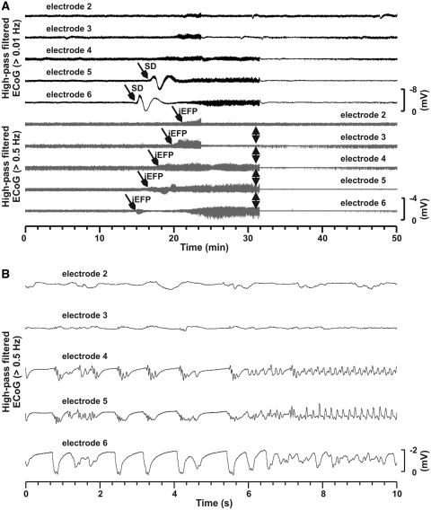
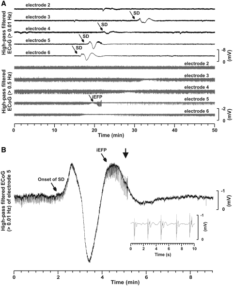
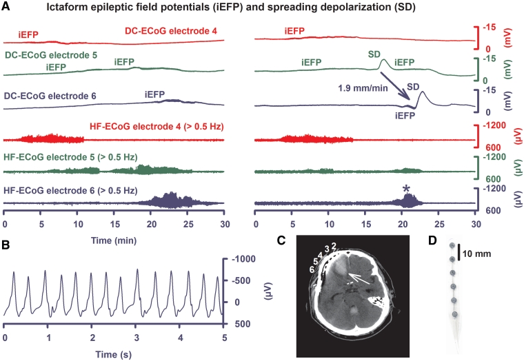
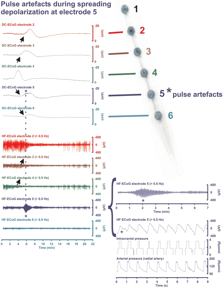
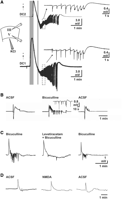
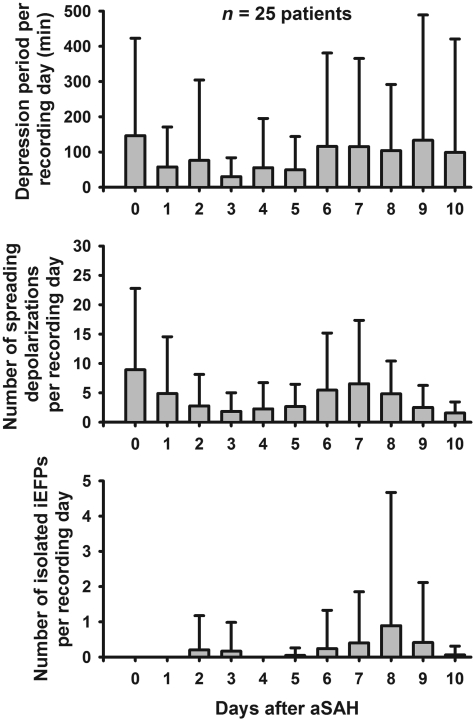
Spreading depolarization of cells in cerebral grey matter is characterized by massive ion translocation, neuronal swelling and large changes in direct current-coupled voltage recording. The near-complete sustained depolarization above the inactivation threshold for action potential generating channels initiates spreading depression of brain activity. In contrast, epileptic seizures show modest ion translocation and sustained depolarization below the inactivation threshold for action potential generating channels. Such modest sustained depolarization allows synchronous, highly frequent neuronal firing; ictal epileptic field potentials being its electrocorticographic and epileptic seizure its clinical correlate. Nevertheless, Leão in 1944 and Van Harreveld and Stamm in 1953 described in animals that silencing of brain activity induced by spreading depolarization changed during minimal electrical stimulations. Eventually, epileptic field potentials were recorded during the period that had originally seen spreading depression of activity. Such spreading convulsions are characterized by epileptic field potentials on the final shoulder of the large slow potential change of spreading depolarization. We here report on such spreading convulsions in monopolar subdural recordings in 2 of 25 consecutive aneurismal subarachnoid haemorrhage patients in vivo and neocortical slices from 12 patients with intractable temporal lobe epilepsy in vitro. The in vitro results suggest that γ-aminobutyric acid-mediated inhibition protects from spreading convulsions. Moreover, we describe arterial pulse artefacts mimicking epileptic field potentials in three patients with subarachnoid haemorrhage that ride on the slow potential peak. Twenty-one of the 25 subarachnoid haemorrhage patients (84%) had 656 spreading depolarizations in contrast to only three patients (12%) with 55 ictal epileptic events isolated from spreading depolarizations. Spreading depolarization frequency and depression periods per 24 h recording episodes showed an early and a delayed peak on Day 7. Patients surviving subarachnoid haemorrhage with poor outcome at 6 months showed significantly higher total and peak numbers of spreading depolarizations and significantly longer total and peak depression periods during the electrocorticographic monitoring than patients with good outcome. In a semi-structured telephone interview 3 years after the initial haemorrhage, 44% of the subarachnoid haemorrhage survivors had developed late post-haemorrhagic seizures requiring anti-convulsant medication. In those patients, peak spreading depolarization number had been significantly higher [15.1 (11.4-30.8) versus 7.0 (0.8-11.2) events per day, P = 0.045]. In summary, monopolar recordings here provided unequivocal evidence of spreading convulsions in patients. Hence, practically all major pathological cortical network events in animals have now been observed in people. Early spreading depolarizations may indicate a risk for late post-haemorrhagic seizures.


Figures

Figure 1
Figure 1
(A) Electrocorticography recording of spreading convulsion. The spreading depolarization started at Electrode 6 55 h after aneurismal SAH and 15 h after discontinuation of the sedation with propofol. It only spread to Electrode 5. The propagation velocity was 5.5 mm/min assuming a wavefront oriented perpendicular to the electrode strip. The peak-to-peak amplitudes were 5.7 and 4.4 mV in Electrodes 6 and 5, respectively. The slow potential change started at Electrode 6 superimposed with ictal epileptic field potentials which propagated from Electrode 6 along the strip until Electrode 2 at an average velocity of 6.6 mm/min. At Electrode 6, two periods with ictal epileptic field potentials were interrupted by a period of silence in brain electrical activity for 3.5 min. The ictal epileptic field potentials showed a maximal amplitude of 2.3 mV and abruptly ceased first in Electrodes 2 and 3 while continuing in Electrodes 4–6 where they simultaneously ceased only 7 min later. The whole ictal epileptic event lasted for 14.5 min. It was followed by complete and long-lasting depression of the brain electrical activity. Recovery of the brain electrical activity started after about 7 min. A clinical seizure was not documented on the intensive care unit. (B) Shows synchronous polyspike wave activity in Electrodes 4–6 at the end of the ictal epileptic event as indicated by the arrows in (A). Note that in this and subsequent figures negative potential deflection is plotted upward.
Figure 2
Figure 2
(A) Electrocorticography recording of spreading convulsion. Two and four hours after the spreading convulsion in Fig. 1, the patient developed two more spreading convulsions but the ictal epileptic field potentials (iEFP) were only observed at Electrode 5 and the spreading depolarization (SD) propagated not only from Electrode 6 to 5 but from 6 to 3 at an average velocity of 2.4 mm/min. (B) Shows in more detail that the ictal epileptic field potential in Electrode 5 started at the end of the slow potential change. The inset demonstrates the polyspike pattern of the ictal epileptic field potentials. Thereafter, the patient had another seven spreading depolarizations propagating from Electrodes 6 to 3 not associated with ictal epileptic field potentials and four ictal epileptic events restricted to Electrode 5, which did not show a close temporal relationship with spreading depolarization. Spreading depolarization and the ictal epileptic activities cleared after Day 4 without anti-convulsant medication. Nevertheless, the patient developed late epilepsy.
Figure 3
Figure 3
Electrocorticography recording showing the cooccurrence of ictal epileptic field potentials with spreading depolarization at the vicinity of a 6 × 2 cm right frontal intracerebral haematoma. (A) On the left, isolated ictal epileptic field potentials are shown spreading from Electrodes 4–6. Such events were clinically associated with short epileptic spells of breath holding and tonic posturing in this unconscious patient. Epileptic ictal activity and seizures disappeared after treatment was started with levetiracetam. On the right, another time period is demonstrated in which ictal epileptic field potentials at Electrode 5 precede and follow the spreading depolarization (spreading convulsion). In the upper three traces the full frequency band from 0 to 45 Hz is given including the direct current component in contrast to Figs 1 and 2 where the lower frequency limit was 0.01 Hz. Full band recordings allow assessment of the slow potential change (indicated by spreading depolarization in the figure) without signal distortion. It is obvious that the slow potential change of spreading depolarization (up to 10.7 mV in the figure) is much larger than that associated with the ictal epileptic field potentials (up to 1.8 mV in the figure). In the lower three traces, a lower frequency limit of 0.5 Hz caused the lower frequency components of the electrocorticography to disappear which eases the assessment of the high-frequency band >0.5 Hz containing the ictal epileptic field potentials. (B) Three per second sharp waves superimposed on shallower delta activity at Electrode 6. In (A), this episode is indicated by the star in the lowest recording trace. (C) CT showing the intracerebral haematoma (white arrow) in the right frontal lobe of the patient and Electrodes 2–6 of the subdural recording strip. Note that both ictal epileptic field potentials and spreading depolarization seem to spread away from the haematoma site. (D) Subdural 6-contact (platinum) electrocorticography recording strip.
Figure 4
Figure 4
Electrocorticography recording of arterial pulse artefacts on the peak of the slow potential change of spreading depolarization at only one electrode (Electrode 5). The upper five traces show the full band recordings from 0 to 45 Hz including the direct current component and thus containing the slow potential change. The lower five traces simultaneously demonstrate the high-frequency (HF) recordings excluding the direct current (DC) component but easing the assessment of the arterial pulse-synchronous electrocorticography changes at Electrode 5 and the spreading depression of activity at Electrodes 2–5. At Electrode 6, the brain electrical activity had been already depressed before the spreading depolarization started. Hence, the spreading depolarization, as indicated by the slow potential change, did not lead to spreading depression of activity here.
Figure 5
Figure 5
Spreading convulsions in human temporal neocortex slices obtained during epilepsy surgery. Spreading depolarizations were induced in these experiments by KCl (2 M) microinjection in layer six. (A) Propagation of the negative direct current (DC) (slow potential) shift characteristic of spreading depolarization from layer five to three followed by ictal epileptic field potentials. In this case, the spreading depolarization triggered the ictal epileptic field potentials spontaneously, i.e. without addition of bicuculline. Note the slow propagation of the spreading depolarization from layer five to three, in contrast to the synchronization of the later ictal burst discharges between the two layers. Lower rows traced by ink writer and upper rows recorded by a digital oscilloscope. (B) The GABAA antagonist bicuculline caused the spreading depolarization to trigger ictal epileptic field potentials in those cases where it did not trigger the ictal epileptic field potentials spontaneously. From left to right: spreading depolarization before, 45 min after bicuculline (5 µM) application and 45 min after washout of bicuculline (lower trace in the middle: digital oscilloscope; upper trace: ink writer). (C) Addition of levetiractem (500 µM) to the bath solution inhibited the induction of ictal epileptic field potentials by the spreading depolarization under continuous application of bicuculline. This effect was reversible. (D) N-methyl-d-aspartate did not cause spreading depolarizations to trigger ictal epileptic field potentials. Spreading depolarization before 45 min, after application of N-methyl-d-aspartate (10 µM) and 45 min after washout.
Figure 6
Figure 6
Temporal distribution of the average depression periods, spreading depolarizations and isolated ictal epileptic events (iEFPs) in the 25 patients over the first 11 days after aneurismal SAH. Whereas the depression periods and spreading depolarizations showed both an early peak on the day of the haemorrhage and a delayed peak on Day 7 after aneurismal SAH, the isolated ictal epileptic events only showed a delayed peak on Day 8. Spreading depolarizations were significantly more frequent than isolated ictal epileptic events on every single day.

Similar articles

Cited by

References

    1. Avoli M, Drapeau C, Louvel J, Pumain R, Olivier A, Villemure JG. Epileptiform activity induced by low extracellular magnesium in the human cortex maintained in vitro. Ann Neurol. 1991;30:589–96. - PubMed
    1. Ayala GF, Dichter M, Gumnit RJ, Matsumoto H, Spencer WA. Genesis of epileptic interictal spikes. New knowledge of cortical feedback systems suggests a neurophysiological explanation of brief paroxysms. Brain Res. 1973;52:1–17. - PubMed
    1. Ayata C, Jin H, Kudo C, Dalkara T, Moskowitz MA. Suppression of cortical spreading depression in migraine prophylaxis. Ann Neurol. 2006;59:652–61. - PubMed
    1. Balestrino M, Young J, Aitken P. Block of (Na+,K+)ATPase with ouabain induces spreading depression-like depolarization in hippocampal slices. Brain Res. 1999;838:37–44. - PubMed
    1. Berger M, Speckmann EJ, Pape HC, Gorji A. Spreading depression enhances human neocortical excitability in vitro. Cephalalgia. 2008;28:558–62. - PubMed

Publication types