. 2011 Nov 27;17(12):1668-73.
doi: 10.1038/nm.2490.

Stimulating healthy tissue regeneration by targeting the 5-HT₂B receptor in chronic liver disease

Affiliations

Stimulating healthy tissue regeneration by targeting the 5-HT₂B receptor in chronic liver disease

Mohammad R Ebrahimkhani et al. Nat Med. .

Abstract

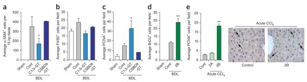
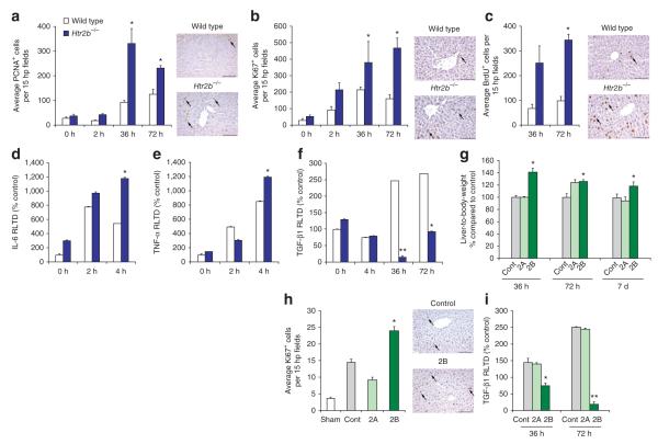
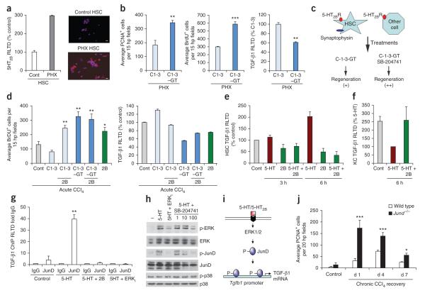
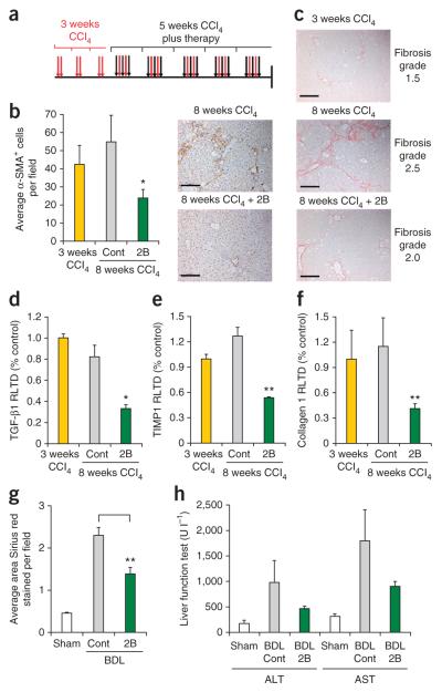
Tissue homeostasis requires an effective, limited wound-healing response to injury. In chronic disease, failure to regenerate parenchymal tissue leads to the replacement of lost cellular mass with a fibrotic matrix. The mechanisms that dictate the balance of cell regeneration and fibrogenesis are not well understood. Here we report that fibrogenic hepatic stellate cells (HSCs) in the liver are negative regulators of hepatocyte regeneration. This negative regulatory function requires stimulation of the 5-hydroxytryptamine 2B receptor (5-HT(2B)) on HSCs by serotonin, which activates expression of transforming growth factor β1 (TGF-β1), a powerful suppressor of hepatocyte proliferation, through signaling by mitogen-activated protein kinase 1 (ERK) and the transcription factor JunD. Selective antagonism of 5-HT(2B) enhanced hepatocyte growth in models of acute and chronic liver injury. We also observed similar effects in mice lacking 5-HT(2B) or JunD or upon selective depletion of HSCs in wild-type mice. Antagonism of 5-HT(2B) attenuated fibrogenesis and improved liver function in disease models in which fibrosis was pre-established and progressive. Pharmacological targeting of 5-HT(2B) is clinically safe in humans and may be therapeutic in chronic liver disease.


Figures

Figure 1
Figure 1
Selective depletion of hepatic stellate cells or antagonism of 5-HT2B stimulates liver growth. (a–c) The average number of α-SMA+ myofibroblasts (a), F4/80+ Kupffer cells (b) and PCNA+ hepatocytes (c) per field, or set of fields, in liver tissue samples from mice that underwent BDL followed by intraperitoneal (i.p.) injections of C1-3–gliotoxin (C1-3–GT), control antibody conjugated to gliotoxin (CSBD9-GT) or saline vehicle (Cont) or from mice that underwent a sham operation without further treatment. (d) Average BrdU-stained hepatocytes per field in liver tissue samples from mice that underwent BDL followed by i.p. injections of the 5-HT2B antagonist SB-204741 (2B) or vehicle (Cont) versus sham operation with no further treatment. (e) Left, the average number of PCNA+ hepatocytes per field in liver tissue samples from mice i.p. injected with CCl4 followed by i.p. administration of vehicle (Cont), 5-HT2A antagonist ketanserin (2A) or the 5-HT2B antagonist SB-204741 (2B). Right, representative images of PCNA-stained liver; arrows denote PCNA+ hepatocytes. Error bars are means ± s.e.m. Data are representative of four or five mice per group. *P < 0.05, **P < 0.01 compared to control, calculated using analysis of variance (ANOVA). Scale bars, 50 μm. Hp, high power.
Figure 2
Figure 2
Gene deletion or blockade of 5-HT2B enhances liver regeneration after PHX. (a–c) Average number of PCNA+ (a), Ki67+ (b) and BrdU+ (c) hepatocytes, per 15 hp fields, in liver sections from wild-type (WT) or Htr2b (5-HT2B receptor)-knockout mice that underwent PHX or a sham operation. Representative images of PCNA, Ki67 and BrDU immunostaining; arrows denote positively stained hepatocytes. (d–f) Whole-liver IL-6 (d), TNF-α (e) and TGF-β1 (f) mRNA levels expressed as relative level of transcription difference (RLTD) at the indicated time points after PHX in WT and knockout mice. (g) Liver-to-body-weight ratio in mice that underwent PHX followed by i.p. injections of vehicle (Cont), ketanserin (2A) or SB-204741 (2B). (h,i) The average number of Ki67+ hepatocytes per 15 hp fields (h) and whole-liver TGF-β1 mRNA levels (i) in mice that underwent PHX followed by i.p. administration of vehicle (Cont), ketanserin (2A), SB-204741(2B) or sham operation. Representative images of Ki67-stained liver; arrows denote Ki67+ hepatocytes. All images are at ×200 magnification; scale bars, 100 μm. Data are means ± s.e.m. and representative of at least four mice per group. Statistical significance was determined by ANOVA, *P < 0.05, **P < 0.01 compared to control, 0 h or 36 h.
Figure 3
Figure 3
5-HT2B blockade enhances liver regeneration by inhibiting induction of TGF-β1 expression by HSC that is dependent on 5-HT, ERK and JunD. (a) Expression of 5-HT2B determined by immunocytochemistry and qRT-PCR (RLTD) in HSCs isolated from the liver of control mice or 36 h PHX mice; n = 3 cell preparations. Scale bars, 10 μm. (b) Average number of PCNA+ and BrDU+ hepatocytes per 15 hp fields and TGF-β1 mRNA levels in mice treated with C1-3 or C1-3–gliotoxin 72 h after PHX. (c) Experiment to determine whether HSCs mediate the regenerative effects of 5-HT2B antagonism. If other cells expressing 5-HT2B are involved, then the combination of C1-3-GT–induced HSC apoptosis and treatment with SB-204741 should generate additive (++) effects compared with C1-3-GT treatment alone (+). (d) Average number of BrdU+ hepatocytes per 15 hp fields and TGF-β1 mRNA levels in livers from mice i.p. injected with CCl4 followed by i.p. administration of either C1-3 or C1-3–gliotoxin ± SB-204741 or vehicle. (e,f) TGF-β1 mRNA levels in mouse HSCs (e) or Kupffer cells (KC) (f) stimulated with 5-HT ± SB-204741. (g) ChIP analysis of JunD recruitment to the TGF-β1 promoter in rat HSCs stimulated with 5-HT for 4 h ± SB-204741 (2B) or ERK inhibitor PD98059 (ERKi). (h) Western blot detection of ERK, phospho-ERK (p-ERK), JunD, phospho-JunD (p-JunD) and p38 or phospho-p38 (p-p38) in rat HSCs that had been pretreated for 30 min with PD98059 or 1–100 μM SB-204741 prior to stimulation with 5-HT. (i) Schematic representation of ERK-dependent recruitment of JunD to the TGF-β1 promoter in HSCs upon 5-HT binding to the 5-HT2B receptor. (j) Average number of PCNA+ hepatocytes in liver sections from WT and JunD-knockout mice at peak injury and recovery (days 1–7 after the final injection) after 8 weeks of CCl4 injury. Data are means ± s.e.m. of at least four mice per group. *P < 0.05, **P < 0.01, ***P < 0.001 compared to control calculated by ANOVA.
Figure 4
Figure 4
Blockade of 5-HT2B receptors attenuates liver fibrosis. (a) Diagram showing the dosing regime in the therapy model; mice were given CCl4 injections i.p. biweekly for 3 weeks, followed by i.p. injections of SB-204741 (2B) or vehicle (Cont) triweekly in addition to biweekly CCl4 for a further 5 weeks. (b) Average number of α-SMA+ cells per hp field and representative images of α-SMA–immunostained liver sections in this model. (c) Fibrosis pathology scoring using the adapted Metavir scale and representative images of livers stained with Sirius red (collagen deposition) in this model. (d–f) Whole-liver TGF-β1, tissue inhibitor of metalloproteinase 1 (TIMP1) and collagen I mRNA levels (RLTD) in this model. (g) Mice underwent BDL, followed by i.p. injections of SB-204741 or vehicle (Cont) daily for 7 d starting at day 7 after BDL. The average Sirius red-positive area per field was calculated using morphometric analysis. (h) Serum transaminases, alanine transaminase (ALT) and aspartate transaminase (AST), markers of liver function, were assessed at 14 d after BDL ± SB-204741 or after sham operation. Photomicrographs are at ×100 magnification. Scale bars, 100 μm. Data are means ± s.e.m. n = 5 mice per group. *P < 0.05, **P < 0.01 compared to control calculated by ANOVA.

Comment in

References

    1. Gurtner GC, Werner S, Barrandon Y, Longaker MT. Wound repair and regeneration. Nature. 2008;453:314–321. - PubMed
    1. Wallace K, Burt AD, Wright MC. Liver fibrosis. Biochem. J. 2008;411:1–18. - PubMed
    1. Marshall A, et al. Relation between hepatocyte G1 arrest, impaired hepatic regeneration, and fibrosis in chronic hepatitis C virus infection. Gastroenterology. 2005;128:33–42. - PubMed
    1. Roskams T. Liver stem cells and their implication in hepatocellular and cholangiocarcinoma. Oncogene. 2006;25:3818–3822. - PubMed
    1. Malik R, Selden C, Hodgson H. The role of non-parenchymal cells in liver growth. Semin. Cell Dev. Biol. 2002;13:425–431. - PubMed

Publication types

MeSH terms