Review
. 2012 Feb;33(1):63-76.
doi: 10.1016/j.mam.2011.11.001. Epub 2011 Nov 22.

Vitamin A and immune regulation: role of retinoic acid in gut-associated dendritic cell education, immune protection and tolerance

Affiliations
Review

Vitamin A and immune regulation: role of retinoic acid in gut-associated dendritic cell education, immune protection and tolerance

Barbara Cassani et al. Mol Aspects Med. 2012 Feb.

Abstract

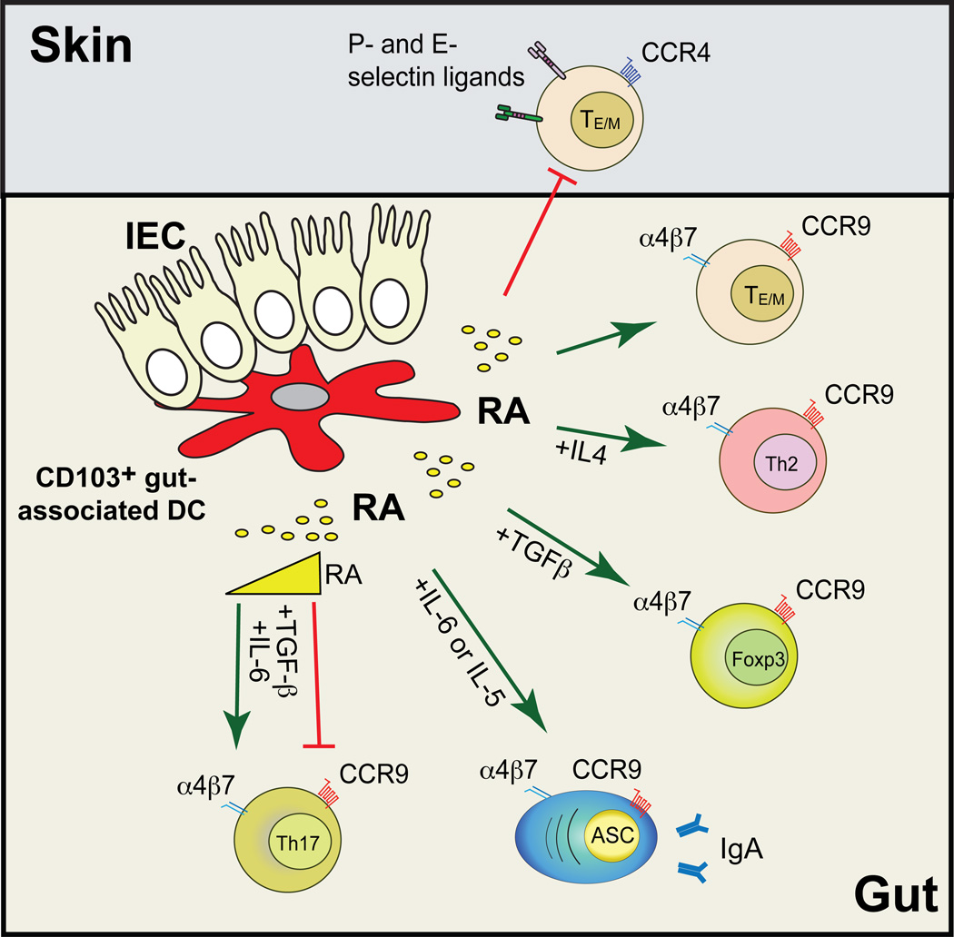
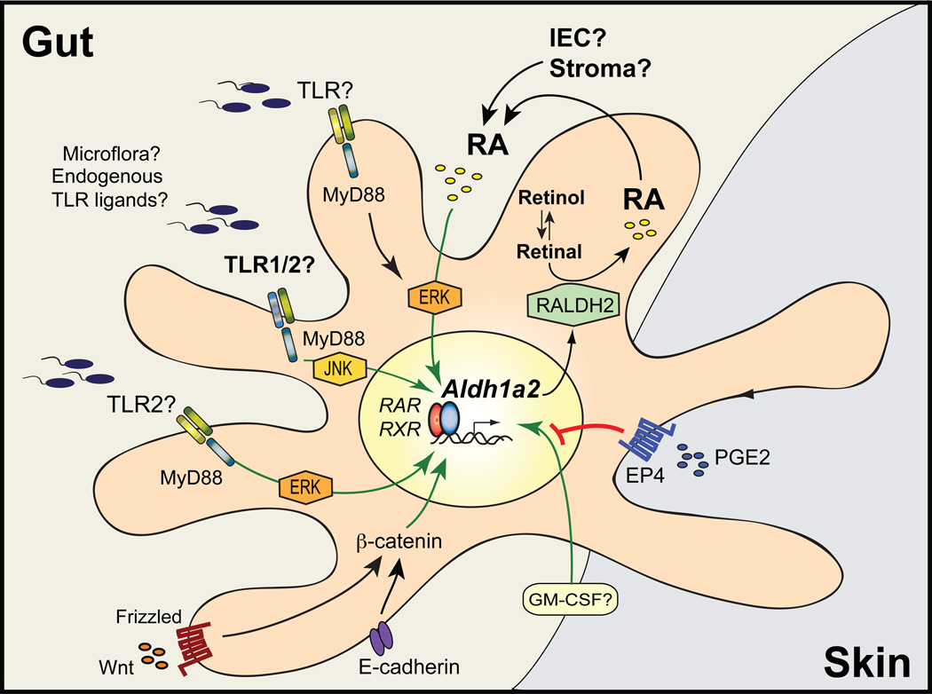
The vitamin A (VA) metabolite all-trans retinoic acid (RA) plays a key role in mucosal immune responses. RA is produced by gut-associated dendritic cells (DC) and is required for generating gut-tropic lymphocytes and IgA-antibody-secreting cells (IgA-ASC). Moreover, RA modulates Foxp3(+) regulatory T cell (T(REG)) and Th17 effector T cell differentiation. Thus, although RA could be used as an effective "mucosal adjuvant" in vaccines, it also appears to be required for establishing intestinal immune tolerance. Here we discuss the roles proposed for RA in shaping intestinal immune responses and tolerance at the gut mucosal interface. We also focus on recent data exploring the mechanisms by which gut-associated DC acquire RA-producing capacity.


Conflict of interest statement

Conflicts of interest: The authors declare no competing financial interests.

Figures

Figure 1
Figure 1. Retinoic acid orchestrates intestinal immunity
CD103+ gut-associated-DC synthesize all-trans retinoic acid (RA), which is necessary and sufficient to induce gut homing receptors α4β7 and CCR9 on effector T and B cells, while reciprocally inhibiting the expression of skin homing receptors (P- and E-selectin ligands and CCR4). RA also potentiates the differentiation of Foxp3+ regulatory T cells (requiring TGF-β) and Th2 cells (requiring Th2 polarizing cytokines). In addition, RA promotes the induction of IgA-ASC (also needing IL-5 and/or IL-6). Of note, RA seems to be required for the induction of intestinal Th17 cells, although medium to high RA concentrations have been shown to inhibit ex vivo Th17 differentiation. It is also unclear whether RA is needed for extra-intestinal Th17 responses. Green and red lines indicate induction or inhibition, respectively.
Figure 2
Figure 2. Mechanisms proposed to induce Aldh1a2 in gut-associated DC
Aldh1a2 is the main RALDH isoform expressed in gut-associated DC, although Aldh1a1 and Aldh1a3 have also been described in other gut-associated cell types, such as IEC and stromal cells. Whereas the mechanisms controlling the expression of the latter RALDH isoforms remain to be determined, recent literature show that the expression of Aldh1a2 in DC can be modulated by several microenvironmental stimuli. In particular, recent evidence indicates that RA itself is necessary and sufficient to induce its own synthesis by upregulating Aldh1a2 in DC as a positive feedback loop. IEC are among the potential sources of RA to educate DC in the gut, although this remains to be demonstrated. In addition, several other factors have been proposed to induce Aldh1a2 in DC, including MyD88-dependent TLR2 signals, Wnt/β-catenin pathway, GM-CSF, and PPARγ-agonists (the latter not shown in the figure). In contrast, PGE2 acting on EP4 receptor inhibits Aldh1a2 induction in skin-associated DC. However, although the critical role of RA in Aldh1a2 induction in gut-associated DC has been consistently shown by several groups, the contribution of other factors in physiological gut-associated DC education and the intracellular signaling pathways mediating these effects (including MyD88, ERK and JNK) are currently less clear. Green and red lines indicate induction or inhibition of Aldh1a2 expression, respectively.

Similar articles

Cited by

References

    1. Acharya M, Mukhopadhyay S, Paidassi H, Jamil T, Chow C, Kissler S, Stuart LM, Hynes RO, Lacy-Hulbert A. alphav Integrin expression by DCs is required for Th17 cell differentiation and development of experimental autoimmune encephalomyelitis in mice. The Journal of clinical investigation. 2010;120(12):4445–4452. - PMC - PubMed
    1. Amsen D, Blander JM, Lee GR, Tanigaki K, Honjo T, Flavell RA. Instruction of distinct CD4 T helper cell fates by different notch ligands on antigen-presenting cells. Cell. 2004;117(4):515–526. - PubMed
    1. Benson MJ, Pino-Lagos K, Rosemblatt M, Noelle RJ. All-trans retinoic acid mediates enhanced T reg cell growth, differentiation, and gut homing in the face of high levels of co-stimulation. J Exp Med. 2007 1775-1774. - PMC - PubMed
    1. Bhat PV. Retinal dehydrogenase gene expression in stomach and small intestine of rats during postnatal development and in vitamin A deficiency. FEBS Lett. 1998;426(2):260–262. - PubMed
    1. Bjersing JL, Telemo E, Dahlgren U, Hanson LA. Loss of ileal IgA+ plasma cells and of CD4+ lymphocytes in ileal Peyer's patches of vitamin A deficient rats. Clin Exp Immunol. 2002;130(3):404–408. - PMC - PubMed

Publication types

MeSH terms