. 2012 Jun 1;520(8):1800-18.
doi: 10.1002/cne.23014.

Bilateral and ipsilateral ascending tectopulvinar pathways in mammals: a study in the squirrel (Spermophilus beecheyi)

Affiliations

Bilateral and ipsilateral ascending tectopulvinar pathways in mammals: a study in the squirrel (Spermophilus beecheyi)

Felipe Fredes et al. J Comp Neurol. .

Abstract

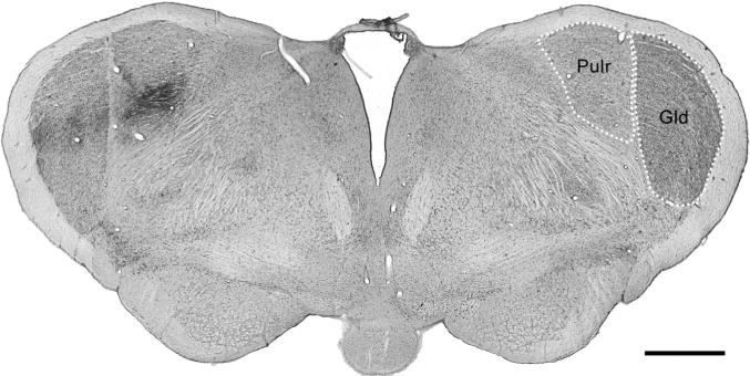
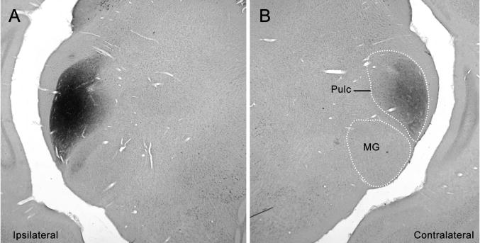
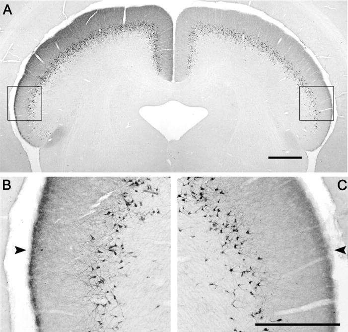
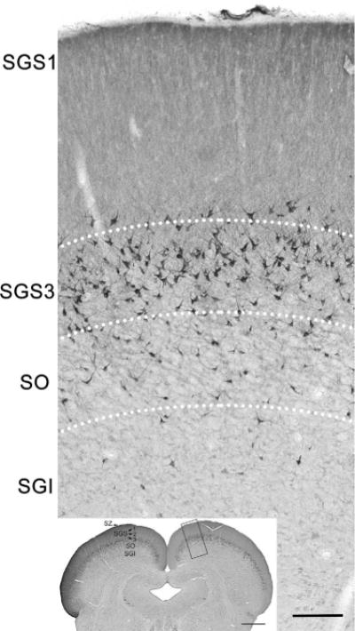
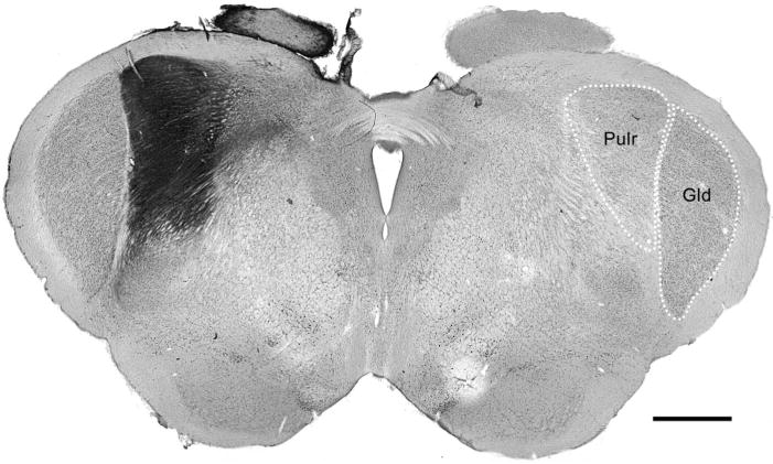
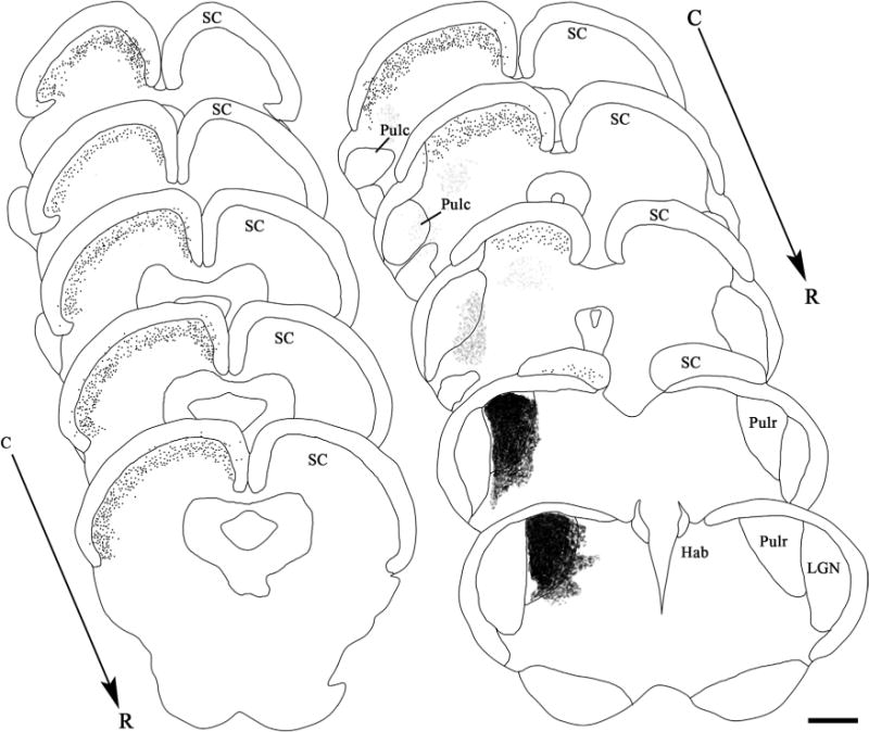
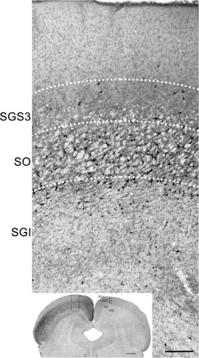
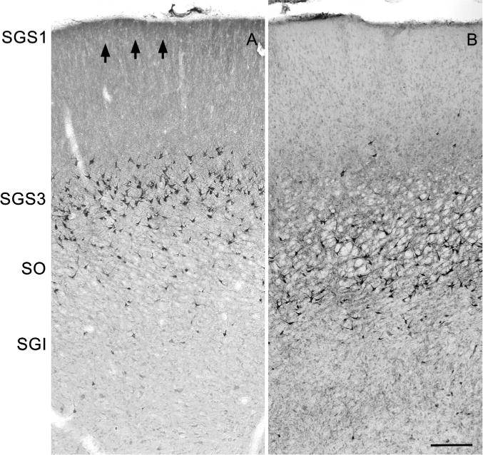
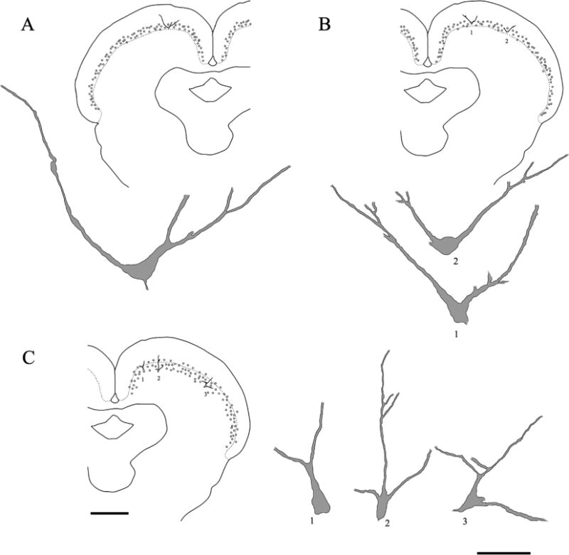
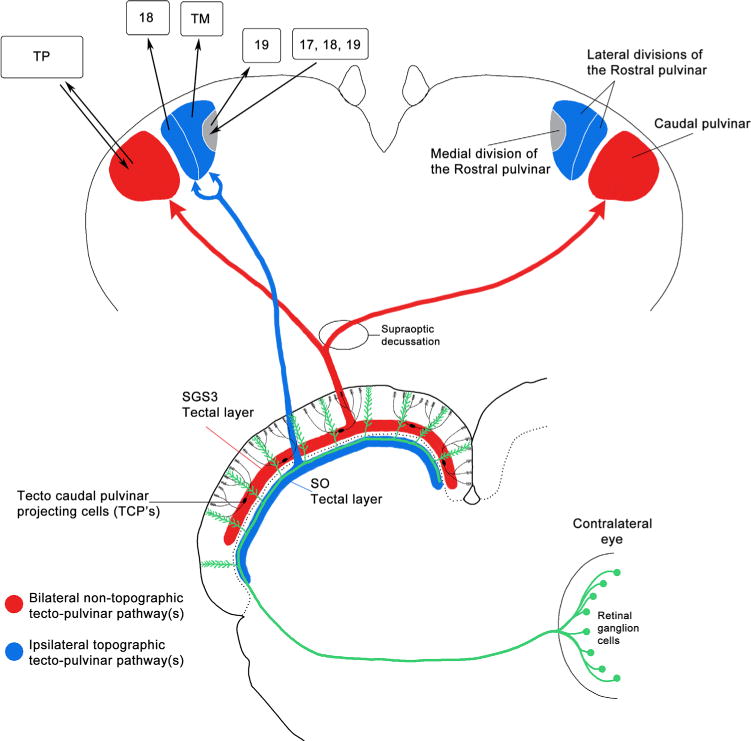
The mammalian pulvinar complex is a collection of dorsal thalamic nuclei related to several visual and integrative processes. Previous studies have shown that the superficial layers of the superior colliculus project to multiple divisions of the pulvinar complex. Although most of these works agree about the existence of an ipsilateral tectopulvinar projection arising from the stratum griseum superficialis, some others report a bilateral projection originating from this same tectal layer. We investigated the organization of the tectopulvinar projections in the Californian ground squirrel using cholera toxin B (CTb). We confirmed previous studies showing that the caudal pulvinar of the squirrel receives a massive bilateral projection originating from a specific cell population located in the superficial collicular layers (SGS3, also called the "lower SGS" or "SGSL"). We found that this projection shares striking structural similarities with the tectorotundal pathway of birds and reptiles. Morphology of the collicular cells originating this projection closely corresponds to that of the bottlebrush tectal cells described previously for chickens and squirrels. In addition, we found that the rostral pulvinar receives an exclusively ipsilateral projection from a spatially separate population of collicular cells located at the base of the stratum opticum, deeper than the cells projecting to the caudal pulvinar. These results strongly support, at a structural level, the homology of the pathway originating in the SGS3 collicular cells upon the caudal pulvinar with the tectorotundal pathway of nonmammalian amniotes and contribute to clarifying the general organization of the tectopulvinar pathways in mammals.


Figures

Figure 1
Figure 1
Anterograde labeling of the rostral nucleus of the pulvinar complex after a CTb injection into the superior colliculus. Microphotographs of CTb-reacted, giemsa-counterstained sections. A: CTb injection in the lateral aspect of the superior colliculus (SC). Approximate SC layering is indicated at the contralateral side. Note that the main deposit of tracer concentrates in the superficial SC layers. B: Anterograde labeling of the rostral pulvinar nucleus (Pulr) and dorsal lateral geniculate (Gld) as a result of the injection shown in A. Note the patchy appearance of the terminals in the ipsilateral rostral pulvinar. Contralateral rostral pulvinar appears free of label. SZ, stratum zonale; SGS, stratum griseum superficiale; SO, stratum opticum; SGI, stratum griseum intermedium. Scale bar = 1 mm.
Figure 2
Figure 2
Serial reconstruction of the pattern of labeled processes resulting from a CTb injection in the superior colliculus (same case as in Figs. 1, 3). Drawings are oriented from caudal (C) to rostral (R). Lighter and darker gray shadowing indicates the intensity of the label. The injection site is represented in solid black. The caudal pulvinar appears bilaterally labeled through all its rostrocaudal extension. NOT, nucleus of the optic tract; Hab, medial habenula; GLv, ventral geniculate nucleus. Other abbreviations as in Figures 1 and 3. Scale bar = 1 mm.
Figure 3
Figure 3
Anterograde labeling of the caudal nucleus of the pulvinar complex as a result of the CTb injection shown in Figure 1. A: CTb-reacted, giemsa-counterstained coronal section at the level of the tectal comissure. Note the bilateral anterograde labeling in the caudal pulvinar (Pulc). B. CTb-reacted-only coronal section at relatively the same level as in A. The labeled processes span the whole extent of the nucleus at both sides, but the ipsilateral side presents a denser plexus of fibers. MG, medial geniculate nucleus. Scale bar = 1 mm.
Figure 4
Figure 4
CTb injection into the deep laminae of the superior colliculus. Microphotographs of CTb-reacted, giemsa-counterstained coronal sections at the level of the injection site (A) and rostral to it (B). The caudal pulvinar appears free of label at both sides. Note the very dense labeling at the ipsilateral nucleus of the optic tract (NOT). Other abbreviations as in Figures 1 and 3. Scale bar = 1 mm.
Figure 5
Figure 5
Anterograde labeling of the rostral nucleus of the pulvinar complex after the CTb injection shown in Figure 4. Microphotograph of a CTb-reacted, giemsa-counterstained coronal section. Only a restricted area of the ipsilateral rostral pulvinar is labeled. A localized plexus of labeled processes is also evident at the ipsilateral dorsal geniculate nucleus. Scale bar = 1 mm.
Figure 6
Figure 6
CTb injection site into the caudal pulvinar and resulting labeling of the contralateral caudal pulvinar. Microphotograph of a CTb-reacted, giemsa-counterstained section. Note that the injection site is restricted to the caudal pulvinar, with no diffusion of tracer to any other contiguous structures (A). At the contralateral caudal pulvinar, a dense plexus of labeled terminals made up a “specular image” of the injection site (B).
Figure 7
Figure 7
Bilateral retrograde labeling of tectal cells after the CTb injection shown in Figure 6. Microphotographs of a CTb-reacted coronal section at the level of the tectal comissure (A). Retrogradely labeled cell bodies can be seen through the whole extent of the colliculus at both sides. Note that the density of labeled cell bodies is greater in the ipsilateral side than in the contralateral one. B,C: Higher magnifications of the boxed areas in A. Note that most of the cells exhibit two or more primary dendrites directed obliquely toward the collicular surface. Note also the superficial band of label indicated by the arrowhead. Scale bars = 1 mm in A; 500 μm in C (applies to B,C).
Figure 8
Figure 8
Serial reconstruction of the pattern of labeled processes resulting from a CTb injection into the caudal pulvinar. Drawings are displayed from caudal (C) to rostral (R). Gray shadowing indicate labeled processes. Black dots indicate labeled cell bodies. Solid black represented the injection site. Note that the labeled processes at the contralateral side form a specular image of the injection site. Note also that the retrograded labeled cell bodies span bilaterally trough all levels of the superior colliculus. NOT, nucleus of the optic tract; Hab, medial habenula; GLv, ventral geniculate nucleus. Other abbreviations as in Figures 1 and 3. Scale bar = 1 mm.
Figure 9
Figure 9
Laminar location of the collicular cells projecting to the caudal pulvinar. Inset: Microphotograph of a CTb-reacted, giemsa-counterstained section at the level of the oculomotor nuclei showing the retrograde labeled cells after the CTb injection shown in Figure 6. Top: Higher magnification photograph of the boxed area in the inset. Most of the labeled cells are located in the SGS3 layer, but some of these cells are located in the SO layer as well. Note also the dense band of at the SGS1. Abbreviations as in Figure 1. Scale bars = 1 mm in inset; 100 μm in top portion.
Figure 10
Figure 10
CTb injection into the rostral pulvinar. Microphotograph of a CTb-reacted, giemsa-counterstained section at the rostral pulvinar level showing the main CTb deposit into the nucleus. Note that there is a minimal diffusion of the tracer to the adjacent medial regions. Abbreviations as in Figure 1. Scale bar = 1 mm.
Figure 11
Figure 11
Serial reconstruction of the pattern of labeled processes and cells resulting from a CTb injection into the rostral pulvinar. Drawings are displayed form caudal (C) to rostral (R). Solid black shading represents the injection site. Gray shading indicates diffusion of the tracer. Black dots represented retrograded labeled cell bodies. Note the cells are present only in the ipsilateral tectal half. No labeling or tracer diffusion can be observed in the caudal pulvinar. Scale bar = 1 mm.
Figure 12
Figure 12
Laminar location of the collicular cells projecting to the rostral pulvinar. Inset: Microphotograph of a CTb-reacted, giemsa-counterstained coronal section at the level of the oculomotor nuclei showing the retrograde labeled cells resulting from the injection shown in Figure 10. Top: Higher magnification photograph of the boxed area in the inset. Most of the labeled cells are located in the SO layer, but some of the cells are located in the upper SGI sublamina as well. Note also that no labeled cells can be seen in the more superficial layers. Abbreviations as in Figure 1. Scale bars = 1 mm in inset; 100 μm in top portion.
Figure 13
Figure 13
Comparison of the laminar location of the tectal cells projecting to the pulvinar complex. High-power photographs of the tectal cells projecting to the caudal (A) and rostral (B) pulvinar. The cells were retrogradely labeled after CTb injections in the corresponding nuclei. Note that the cells projecting to the caudal pulvinar are located mainly in the SGS3 layer, although some cells can also be seen in the SO layer. In contrast, most of the cells projecting to the rostral pulvinar are located in the SO layer (only very few of them appear located in the SGI layer). Note also the band of labeling at the SGS1 above the cells projecting to the caudal pulvinar (arrows). Scale bar = 100 μm.
Figure 14
Figure 14
Camera lucida reconstruction of tectal cells projecting to the pulvinar. A: Camera lucida reconstruction of a cell retrogradely labeled after a CTb injection in the ipsilateral caudal pulvinar. B: Camera lucida reconstruction of two cells retrogradely labeled after a CTb injection in the contralateral caudal pulvinar. Note that, in both cases, cells show two primary dendrites running oblique to the tectal surface. C: Camera lucida reconstructions of three cells retrogradely labeled after a CTb injection into the ipsilateral rostral pulvinar. Note the more fusiform shape and the smaller size of these cells compared with the cells depicted in A and B. The relative positions of the reconstructed cells are indicated within the corresponding tectal schema. Circles indicates the relative position of all retrogradely labeled cells. They do not represent actual numbers. Scale bars = 1 mm at left (applies to all tectal schema); 50 μm at right (applies to all reconstructed cells).
Figure 15
Figure 15
Summary of the organization of the tectopulvinar pathways in the squirrel. Two parallel pathways arise from different tectal populations. Tectal ganglion cells with bottlebrush dendritic endings (black; Major et al., 2000) located in the layer three of the stratum griseum superficiale 3 project bilaterally to the caudal pulvinar in a nontopographic manner (red). The contralateral projection arises from axonal collaterals that cross the midline by the supraoptic decussation. A different population of cells, located in the lower margin of the stratum opticum (SO), projects exclusively ipsilaterally to the lateral subdivisions of the rostral pulvinar. The medial division of the rostral pulvinar is devoid of tectal afferents (gray; Robson and Hall, 1977; Baldwin et al., 2011). At upper left, the cortical connectivity of the pulvinar divisions is shown based on the literature (Kaas et al., 1972; Robson and Hall, 1977; Wong et al., 2008). Most of the retinal ganglion cells (green) project topographically to the contralateral superior colliculus entering by the SO tectal layer with terminations in the three layers of the stratum griseum superficiale (Major et al., 2003). Note that the bottlebrush dendritic endings of the caudal pulvinar projecting cells colocalize with the retinal terminals in the stratum griseum superficiale 1.

Similar articles

Cited by

References

    1. Aboitiz F. Genetic and developmental homology in amniote brains. Toward conciliating radical views of brain evolution. Brain Res Bull. 2011;84:125–136. - PubMed
    1. Aboitiz F, Montiel J. Origin and evolution of the vertebrate telencephalon, with special reference to the mammalian neocortex. Adv Anat Embryol Cell Biol. 2007;193:1–112. - PubMed
    1. Aboitiz F, Morales D, Montiel J. The evolutionary origin of the mammalian isocortex: towards an integrated developmental and functional approach. Behav Brain Sci. 2003;26:535–552. discussion 552–585. - PubMed
    1. Abramson BP, Chalupa LM. Multiple pathways from the superior colliculus to the extrageniculate visual thalamus of the cat. J Comp Neurol. 1988;271:397–418. - PubMed
    1. Albano JE, Norton TT, Hall WC. Laminar origin of projections from the superficial layers of the superior colliculus in the tree shrew, Tupaia glis. Brain Res. 1979;173:1–11. - PubMed

Publication types