Review
. 2012 Mar 15;519(2):118-30.
doi: 10.1016/j.abb.2011.11.015. Epub 2011 Nov 19.

Regulation of the structure and activity of pyruvate carboxylase by acetyl CoA

Affiliations
Review

Regulation of the structure and activity of pyruvate carboxylase by acetyl CoA

Abdussalam Adina-Zada et al. Arch Biochem Biophys. .

Abstract

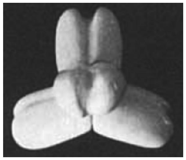
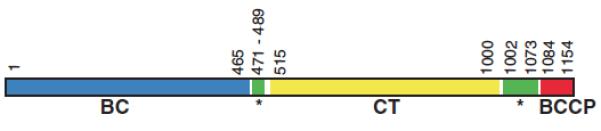
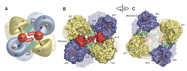
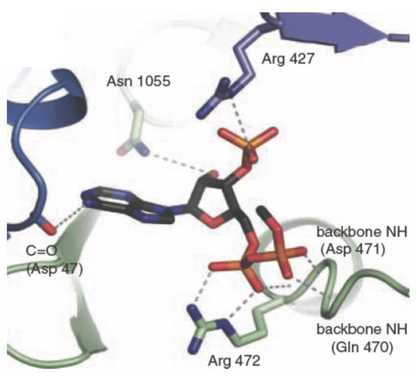
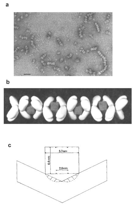
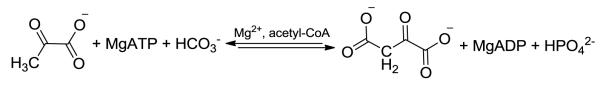
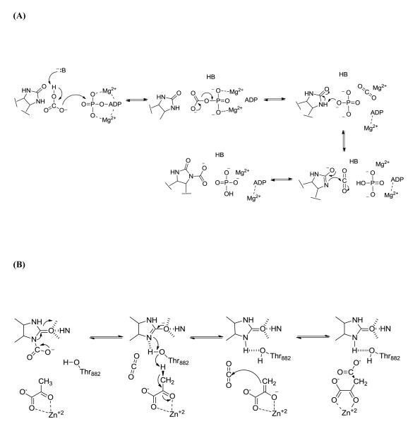
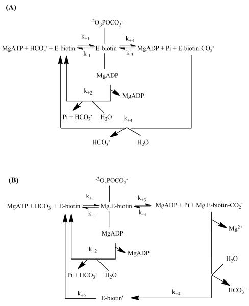
In this review we examine the effects of the allosteric activator, acetyl CoA on both the structure and catalytic activities of pyruvate carboxylase. We describe how the binding of acetyl CoA produces gross changes to the quaternary and tertiary structures of the enzyme that are visible in the electron microscope. These changes serve to stabilize the tetrameric structure of the enzyme. The main locus of activation of the enzyme by acetyl CoA is the biotin carboxylation domain of the enzyme where ATP-cleavage and carboxylation of the biotin prosthetic group occur. As well as enhancing reaction rates, acetyl CoA also enhances the binding of some substrates, especially HCO3-, and there is also a complex interaction with the binding of the cofactor Mg2. The activation of pyruvate carboxylase by acetyl CoA is generally a cooperative processes, although there is a large degree of variability in the degree of cooperativity exhibited by the enzyme from different organisms. The X-ray crystallographic holoenzyme structures of pyruvate carboxylases from Rhizobium etli and Staphylococcus aureus have shown the allosteric acetyl CoA binding domain to be located at the interfaces of the biotin carboxylation and carboxyl transfer and the carboxyl transfer and biotin carboxyl carrier protein domains.


Figures

Figure 1
Figure 1
Model of tetrameric structure of chicken and sheep PC based on electron micrographs of the enzymes. Reproduced with permission from Mayer et al. [57].
Figure 2
Figure 2
Schematic of the domain structure of R. etli PC relative to the amino acid sequence of the enzyme. Reproduced from St. Maurice et al. [9].
Figure 3
Figure 3
(A) Model of the R. etli PC tetramer showing the movement of the BCCP domain between neighboring active sites on opposing polypeptide chains. (B) Surface representation of the top face of the tetramer. For clarity, one of the two individual monomers has been outlined in black. The distance between ATP-γ-S in the BC active site and Zn2+ in the CT active site of the opposing polypeptide chain is 65 Å. (C) Surface representation of the bottom face of the tetramer, after a 180° rotation about the y axis. The BCCP domain is disordered in these monomers and could not be modeled. The distance between ATP-γ-S in the BC active site and Zn2+ in the CT active site of the opposing polypeptide chain increases to 80 Å as a result of the altered orientation of the BC domain. Reproduced from St. Maurice et al. [9].
Figure 4
Figure 4
Allosteric binding site of R. etli PC showing bound ethyl CoA. Reproduced from St. Maurice et al. [9].
Figure 5
Figure 5
a. Electron micrograph of complexes of chicken PC and avidin when the ratio of enzyme: avidin was 2:1, note the many chain-like complexes. The bar represents 40 nm. b. Model of a chain-like complex between PC and avidin, showing how the box-like avidin molecules with their pairs of biotin-binding sites on opposite faces of the box act to “glue” together pairs of subunits from two different PC tetramers. c. Schematic showing a side view of the interaction between a pair of biotin-binding sites on an avidin molecule and the biotins from a pair of PC subunits and the restricted conformation of the subunits to enable this to occur. Figures reproduced with permission from Johannssen et al. [74].
Scheme 1
Scheme 1
The overall pyruvate carboxylation reaction catalysed by PC.
Scheme 2
Scheme 2
Pyruvate carboxylation occurs in two distinct steps. (A) The MgATP-dependent carboxylation of the tethered biotin cofactor occurs in the biotin carboxylase domain. After carboxylation, carboxybiotin it translocated to the carboxyl transferase domain (B) where pyruvate is carboxylated to form oxaloacetate.
Scheme 3
Scheme 3
Reaction schemes for ATP cleavage and biotin carboxylation in (A) chicken PC and (B) Pyc1 proposed on the basis of pre-steady state analysis of these reactions. Reproduced with permission from (A) Legge et al. [49], (B) Branson et al. [46].

References

    1. Jitrapakdee S, Wallace JC. Biochem. J. 1999;340:1–16. - PMC - PubMed
    1. Jitrapakdee S, Wutthisathapornchai A, Wallace JC, MacDonald MJ. Diabetologia. 2010;53:1019–1032. - PMC - PubMed
    1. Jitrapakdee S, Vidal-Puig A, Wallace JC. Cell. Mol. Life. Sci. 2006;63:843–854. - PMC - PubMed
    1. Jitrapakdee S, St. Maurice M, Rayment I, Cleland WW, Wallace JC, Attwood PV. Biochem. J. 2008;413:369–387. - PMC - PubMed
    1. Mukhopadhyay B, Patel VJ, Wolfe RS. Arch. Microbiol. 2000;174:406–414. - PubMed

Publication types