. 2011 Nov 27;18(12):1331-1335.
doi: 10.1038/nsmb.2189.

Exploiting oncogene-induced replicative stress for the selective killing of Myc-driven tumors

Affiliations

Exploiting oncogene-induced replicative stress for the selective killing of Myc-driven tumors

Matilde Murga et al. Nat Struct Mol Biol. .

Abstract

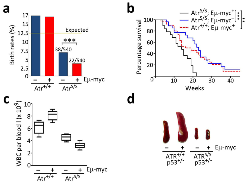
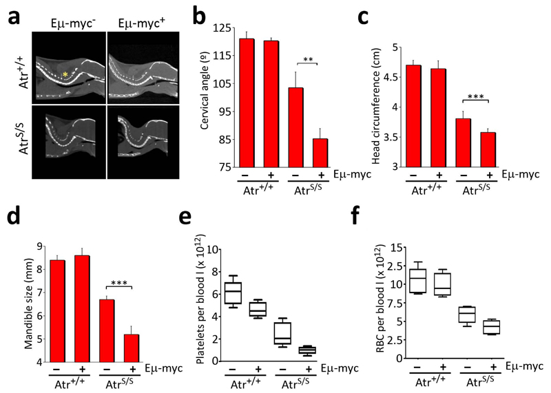
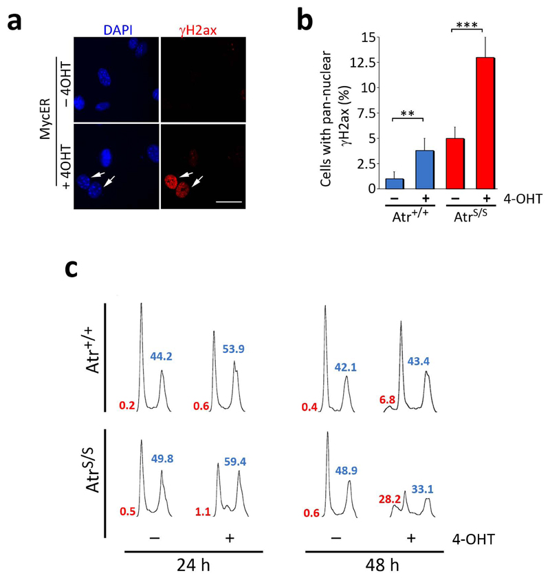
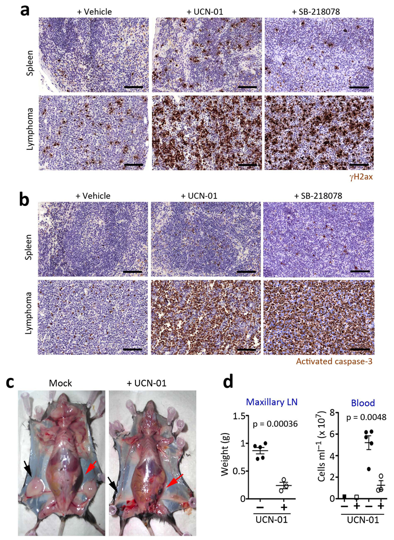
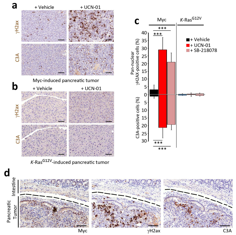
Oncogene-induced replicative stress activates an Atr- and Chk1-dependent response, which has been proposed to be widespread in tumors. We explored whether the presence of replicative stress could be exploited for the selective elimination of cancer cells. To this end, we evaluated the impact of targeting the replicative stress-response on cancer development. In mice (Mus musculus), the reduced levels of Atr found on a mouse model of the Atr-Seckel syndrome completely prevented the development of Myc-induced lymphomas or pancreatic tumors, both of which showed abundant levels of replicative stress. Moreover, Chk1 inhibitors were highly effective in killing Myc-driven lymphomas. By contrast, pancreatic adenocarcinomas initiated by K-Ras(G12V) showed no detectable evidence of replicative stress and were nonresponsive to this therapy. Besides its impact on cancer, Myc overexpression aggravated the phenotypes of Atr-Seckel mice, revealing that oncogenes can modulate the severity of replicative stress-associated diseases.


Figures

Fig. 1
Fig. 1
Reduced levels of Atr prevent lymphomagenesis on Eμ-myc mice. (a) Observed birth rates of the different genotypes obtained from Atr+/S; Eμ-myc+ x Atr+/S; Eμ-myc- crosses (n = 512 mice; χ2: P<0.001). (b) Kaplan Meyer analysis of the indicated genotypes (AtrS/S; Eμ-myc+: 24; Atr+/+; Eμ-myc+: 31; AtrS/S; Eμ-myc-: 32). The asterisks indicate the statistical significance of the different survival curves obtained in the Log- Rank Mantel-Cox Test. There was no significant difference between the survival of AtrS/S and Eμ-myc+ single mutants (p=0.48). (c) White blood cell (WBC) counts from 2 old month mice without detectable lymphoma (n=5). Note the expansion of the WBC compartment on Atr+/+; Eμ-myc+ mice, which contrasts with the further depletion of WBC when the Eμ-myc transgene is combined with Atr hypomorphism. (d) Photograph of the spleens of 5-week old mice illustrating the absence of splenomegalia on AtrS/S; Eμ-myc+; p53+/- mice. (*: P<0.05; **: P<0.01; ***: P<0.001)
Fig. 2
Fig. 2
Eμ-myc aggravates the symptoms of the Seckel Syndrome. (a) Representative images of the computerized tomography (CT) analysis of the spine, illustrating the angle used for the cervical angle measurements (yellow asterisk). (b) Measurements made from (a). (c) Head circumference and (d) right mandible size from 2.5 month old mice of the indicated genotypes were calculated from CT analysis of the crania. (n=6; **: P<0.01; ***: P<0.001) (e) Platelet and (f) red-blood cell (RBC) counts from 2 old month mice without detectable lymphoma (n=5).
Fig. 3
Fig. 3
Atr suppresses Myc-induced RS and apoptosis. (a) Images illustrating the staining pattern of the γH2ax (red) signal on MycER infected MEF. Scale bar indicates 10 μm. (b) Percentage of cells showing a pan-nuclear distribution of γH2ax in MycER infected MEF of the different genotypes in the presence or absence of 4-OHT for 24 hrs. (c) Cell cycle profiles of MycER infected MEF of the different genotypes in the presence or absence of 4-OHT for 24 or 48 hrs. Sub-G1 (red) and S+G2 (blue) percentages are indicated. The images show a representative experiment that was repeated 3 times with independent MEF pairs. (**: P<0.01; ***: P<0.001)
Fig. 4
Fig. 4
Treatment of Myc-induced lymphomas with Chk1 inhibitors. Representative images of (a) γH2ax and (b) activated caspase-3 (C3A) immunohistochemistry (IHC) done on the spleens of mice with or without Myc-induced lymphomas, after a 16 hr treatment with UCN-01 or SB-218078 (5 mg per kg). Black scale bars indicate 100 μm. (c) Representative images of mice carrying Myc-induced lymphomas with or without a daily treatment of UCN-01 (5 mg per kg) for 9 days. The arrows indicate the enlarged lymph nodes (black) and spleen (red) present in the mice carrying Myc-induced lymphomas to illustrate the regression of the tumor upon treatment. (d) Weights of the maxillary lymph nodes and total blood cell counts from the mice mentioned in (c).
Fig. 5
Fig. 5
Effect of Chk1 inhibitors in Myc- or Ras-driven pancreatic tumors. Representative images of γH2ax and activated caspase-3 (C3A) IHC done on pancreatic tumors obtained from (a) Ela-myc or (b) K-RasG12V-knockin mice, after 3 days of treatment with UCN-01 (i.p. 5 mg per kg). Black scale bars indicate 100 μm. (c) Quantification from the analysis presented in (a,b). (d) γH2ax, C3A and c-Myc IHC on the pancreatic tumor from an Ela-myc mouse after a 3 day treatment with UCN-01 (i.p. 5 mg per kg). A dashed black line separates the tumoral area from the neighbouring intestine. Black scale bars indicate 100 μm.

Comment in

References

    1. de Bono JS, Ashworth A. Translating cancer research into targeted therapeutics. Nature. 2010;467:543–549. - PubMed
    1. Bryant HE, et al. Specific killing of BRCA2-deficient tumours with inhibitors of poly(ADP-ribose) polymerase. Nature. 2005;434:913–917. - PubMed
    1. Farmer H, et al. Targeting the DNA repair defect in BRCA mutant cells as a therapeutic strategy. Nature. 2005;434:917–921. - PubMed
    1. Luo J, Solimini NL, Elledge SJ. Principles of cancer therapy: oncogene and non-oncogene addiction. Cell. 2009;136 - PMC - PubMed
    1. Raj L, et al. Selective killing of cancer cells by a small molecule targeting the stress response to ROS. Nature. 2011;475:231–234. - PMC - PubMed

Publication types

MeSH terms