. 2012 Aug 9;31(32):3721-32.
doi: 10.1038/onc.2011.534. Epub 2011 Nov 28.

Abnormal MDMX degradation in tumor cells due to ARF deficiency

Affiliations

Abnormal MDMX degradation in tumor cells due to ARF deficiency

X Li et al. Oncogene. .

Abstract

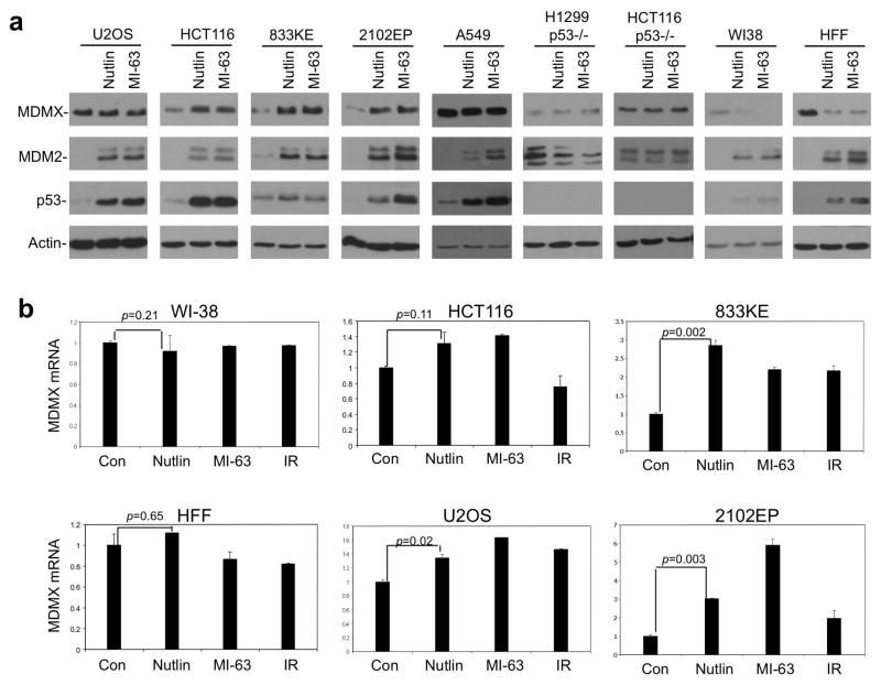
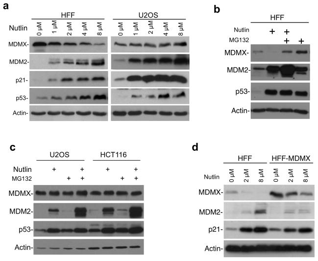
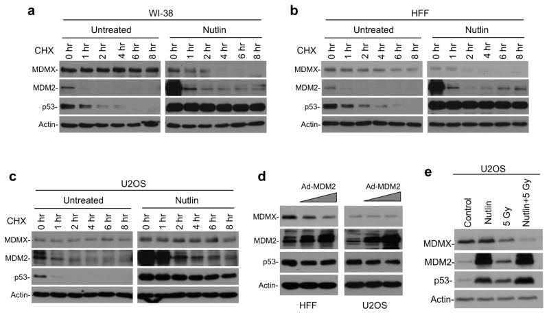
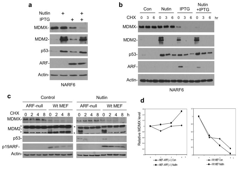
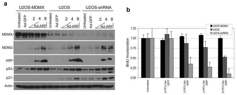
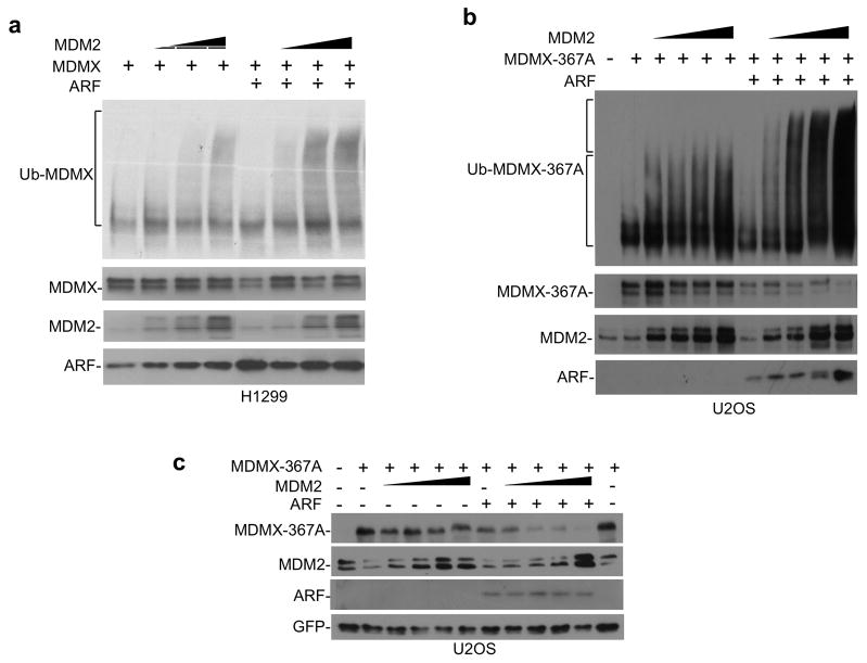
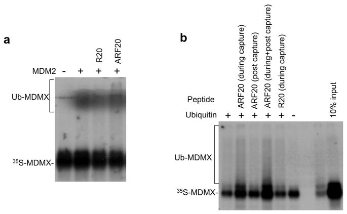
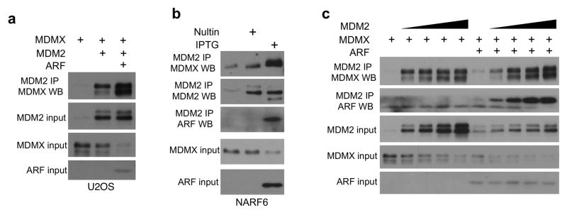
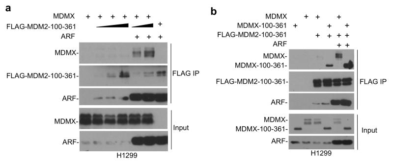
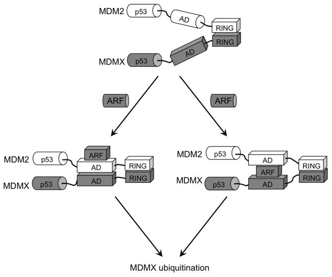
MDMX is a heterodimeric partner of MDM2 and a critical regulator of p53. The MDMX level is generally elevated in tumors with wild-type p53 and contributes to p53 inactivation. MDMX degradation is controlled in part by MDM2-mediated ubiquitination. Here, we show that MDMX turnover is highly responsive to changes in MDM2 level in non-transformed cells, but not in tumor cells. We found that loss of alternate reading frame (ARF) expression, which occurs in most tumors with wild-type p53, significantly reduces MDMX sensitivity to MDM2. Restoration of ARF expression in tumor cells enables MDM2 to degrade MDMX in a dose-dependent manner. ARF binds to MDM2 and stimulates a second-site interaction between the central region of MDM2 and MDMX, and thus increases MDMX-MDM2 binding and MDMX ubiquitination. These results reveal an important abnormality in the p53-regulatory pathway as a consequence of ARF deficiency. Loss of ARF during tumor development not only prevents p53 stabilization by proliferative stress but also causes accumulation of MDMX that compromises p53 activity. This phenomenon may reduce the clinical efficacy of MDM2-specific inhibitors by preventing MDMX downregulation.


Conflict of interest statement

Conflict of interest

The authors declare no conflict of interest.

Figures

Figure 1
Figure 1. MDM2 inhibitors have different effects on MDMX level in normal and tumor cells
(a) Indicated cell lines were treated with 8 μM Nutlin or 8 μM MI-63 for 16 hrs. MDMX level was analyzed by Western blot. WI-38 and HFF are used to represent normal cells, whereas U2OS, HCT116, A549, 833KE and 2102EP are tumor cells. (b) Cells were treated with 8 μM Nutlin, 8 μM MI-63 for 16 hrs or with 10 Gy gamma radiation for 4 hrs. MDMX mRNA level was analyzed by qRT-PCR (n=3).
Figure 2
Figure 2. Proteasome-mediated MDMX degradation after MDM2 induction in normal cells
(a) HFF and U2OS cells were treated with Nultin at indicated concentrations for 16 hrs. MDMX level was analyzed by Western blot. (b) HFF cells were treated with 8 μM Nutlin for 16 hrs, with or without 30 μM MG132 for the last 4 hrs and analyzed by Western blot. (c) U2OS and HCT116 cells were treated with 8 μM Nutlin for 16 hrs, with or without 30 μM MG132 for the last 4 hrs and analyzed by Western blot. (d) Lentivirus-MDMX was used to stably infect HFF to increase its MDMX level. HFF and HFF-MDMX cells were treated with Nutlin for 16 hrs and MDMX level was analyzed by Western blot.
Figure 3
Figure 3. Accelerated MDMX degradation after MDM2 induction in normal cells
(a, b, c) Indicated cell lines were treated with 8 μM Nutlin for 16 hrs. Cells were harvested at different time points after 50 μg/ml CHX was added. MDMX degradation was analyzed by Western blot. (d) HFF and U2OS cells were infected for 24 hrs with MDM2 adenovirus at titers (MOI ranging from 45 to 135 and from 15 to 45, respectively) that produce similar levels of MDM2 and analyzed by Western blot. (e) U2OS cells were treated with 8 μM Nutlin for 16 hrs, followed by 10 Gy IR for 4 hrs, and analyzed for MDMX level.
Figure 4
Figure 4. ARF expression promotes MDMX degradation
(a) NARF6 cells were treated with 100 μM IPTG for 48 hrs to induce ARF. The cells were then treated with 8 μM Nutlin for 16 hrs and analyzed by Western blot. (b) NARF6 cells were treated with IPTG and Nutlin for 24 hrs. CHX (50 μg/ml) were added and cells were harvested at indicated time points for western blot analysis of MDMX stability. (c) Wild type MEF and ARF-null MEF were treated with 8 μM Nutlin for 16 hrs. Cells were harvested at different time points after CHX was added. MDMX level was analyzed by Western blot. Two background bands in the mouse MDM2 blot are marked by *. (d) Quantitation of MDMX levels in (c) by densitometry.
Figure 5
Figure 5. MDMX level has significant impact on ARF-mediated growth arrest
(a) U2OS cells stably transfected with MDMX cDNA (U2OS-MDMX) or MDMX shRNA (U2OS-shRNA) expression plasmids were infected with adenovirus-ARF at indicated MOI and analyzed by western blot after 24 hrs. (b) Cells with MDMX overexpression or knockdown were infected with adenovirus ARF for 24 hrs. Cells were labeled with BrdU for 2 hrs (n=4) and analyzed for DNA replication rate using a BrdU detection ELISA kit.
Figure 6
Figure 6. ARF stimulates MDMX ubiquitination and degradation by MDM2
(a) MDMX was co-transfected with MDM2, ARF and His6-ubiquitin into H1299 cells. Ubiquitinated MDMX was detected by Ni-NTA pull down followed by MDMX Western blot. (b) MDMX-367A was co-transfected with MDM2, ARF and His6-ubiquitin into U2OS cells. Ubiquitinated MDMX-367A was detected by Ni-NTA pull down followed by MDMX Western blot. Note the increase of poly ubiquitinated MDMX in the presence of ARF (upper bracket). (c) MDMX-367A was co-transfected with MDM2 and ARF into U2OS cells. MDMX-367A degradation was analyzed by Western blot.
Figure 7
Figure 7. ARF stimulates MDMX ubiquitination in vitro
(a) An ARF N terminal 20 amino acid peptide (ARF20) or a scrambled peptide (R20) were incubated with recombinant His6-MDM2 and in vitro translated MDMX at 37 °C for 30 min followed by incubation with E1, E2 and ubiquitin for 1 hr. MDMX ubiquitination was detected by SDS-PAGE and autofluorography. (b) SJSA cells were treated with Nutlin for 16 hrs. MDM2 was captured on protein A beads using 4B2 antibody. The beads were used to capture in vitro translated MDMX, followed by washing and incubation with E1, E2 and ubiquitin. ARF20 and R20 peptides were added during MDMX capture, or during incubation with E1/E2/ubiquitin. MDMX binding and ubiquitination was analyzed by SDS-PAGE and autofluorography.
Figure 8
Figure 8. ARF stimulates MDMX-MDM2 binding
(a) MDMX was co-transfected with MDM2 and ARF in U2OS cells. Co-precipitation of MDM2 and MDMX was analyzed by MDM2 IP followed by MDMX Western blot. (b) NARF6 cells were treated with IPTG (to induce ARF) or Nutlin (to induce comparable level of MDM2) for 16 hrs and analyzed for endogenous MDMX-MDM2 binding by IP-Western blot. (c) MDMX was co-transfected with MDM2 and ARF into U2OS cells. MG132 was added 4 hrs before the cells were harvested. Co-precipitation of MDM2 and MDMX was analyzed by MDM2 IP followed by MDMX Western blot.
Figure 9
Figure 9. ARF initiates binding between MDM2 and MDMX acidic domains
(a) FLAG-tagged central region of MDM2 (100–361 out of 1–491) was coexpressed with MDMX and ARF in H1299 cells. MDM2-100–361 binding to full length MDMX was determined by FLAG IP-MDMX Western blot. (b) FLAG-MDM2-100–361 was coexpressed in H1299 cells with MDMX central region (100–361 out of 1–490) and ARF. Binding between MDM2 and MDMX fragments were determined by FLAG IP-MDMX Western blot.
Figure 10
Figure 10. A model of ARF-stimulated MDM2-MDMX heterodimer formation
MDM2 and MDMX C terminal RING domains have intrinsic ability to heterodimerize. ARF binds specifically to MDM2 acidic region, causing conformational change and initiates binding to MDMX central domain. ARF may promote direct MDMX-MDM2 contact (left branch) or act as mediator by binding both MDM2 and MDMX (right branch). The second-site interaction between the central regions increases the stability of MDM2-MDMX complex and promotes MDMX ubiquitination.

Similar articles

Cited by

References

    1. Badciong JC, Haas AL. MdmX is a RING finger ubiquitin ligase capable of synergistically enhancing Mdm2 ubiquitination. J Biol Chem. 2002;277:49668–49675. - PubMed
    1. Bothner B, Lewis WS, DiGiammarino EL, Weber JD, Bothner SJ, Kriwacki RW. Defining the molecular basis of Arf and Hdm2 interactions. J Mol Biol. 2001;314:263–277. - PubMed
    1. Chen D, Shan J, Zhu WG, Qin J, Gu W. Transcription-independent ARF regulation in oncogenic stress-mediated p53 responses. Nature. 2010;464:624–627. - PMC - PubMed
    1. Chen J, Marechal V, Levine AJ. Mapping of the p53 and mdm-2 interaction domains. Mol Cell Biol. 1993;13:4107–4114. - PMC - PubMed
    1. Chen L, Gilkes DM, Pan Y, Lane WS, Chen J. ATM and Chk2-dependent phosphorylation of MDMX contribute to p53 activation after DNA damage. Embo J. 2005;24:3411–3422. - PMC - PubMed

Publication types

MeSH terms