. 2012 Aug 9;31(32):3655-66.
doi: 10.1038/onc.2011.532. Epub 2011 Nov 28.

Regulation of lipid binding underlies the activation mechanism of class IA PI3-kinases

Affiliations

Regulation of lipid binding underlies the activation mechanism of class IA PI3-kinases

W-C Hon et al. Oncogene. .

Abstract

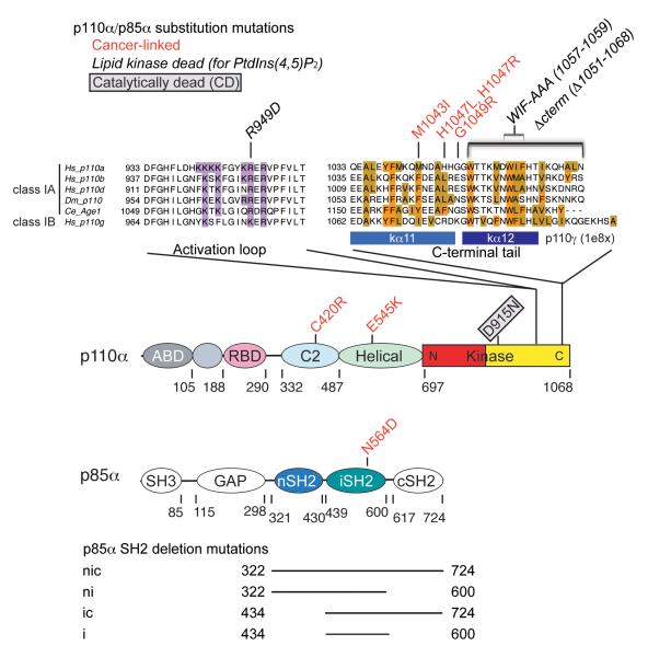
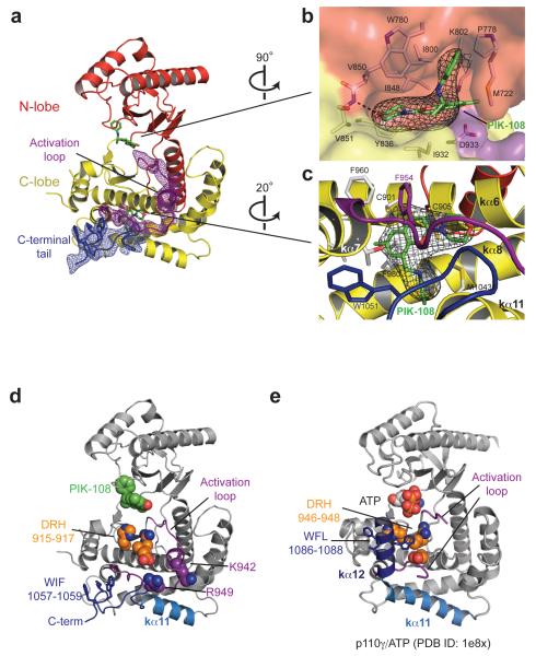
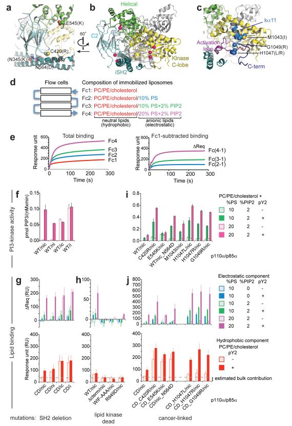
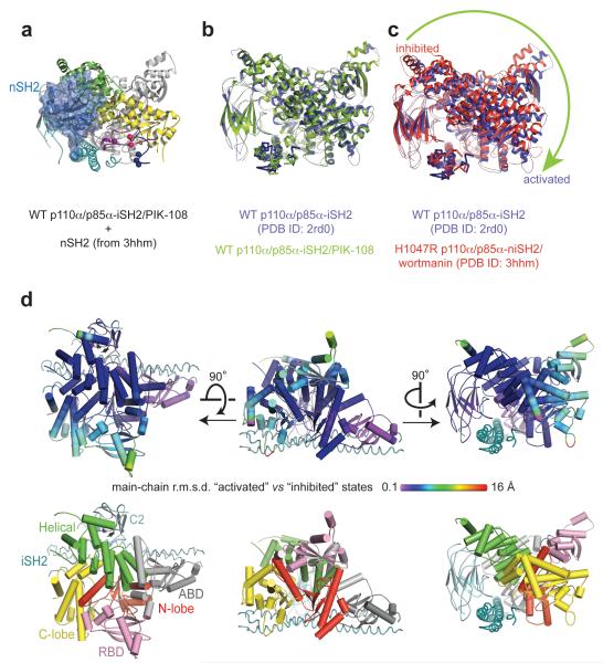
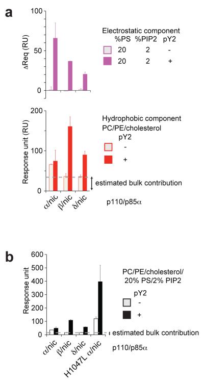
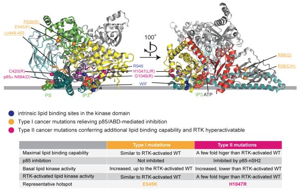
Somatic missense mutations in PIK3CA, which encodes the p110α catalytic subunit of phosphoinositide 3-kinases, occur frequently in human cancers. Activating mutations spread across multiple domains, some of which are located at inhibitory contact sites formed with the regulatory subunit p85α. PIK3R1, which encodes p85α, also has activating somatic mutations. We find a strong correlation between lipid kinase and lipid-binding activities for both wild-type (WT) and a representative set of oncogenic mutant complexes of p110α/p85α. Lipid binding involves both electrostatic and hydrophobic interactions. Activation caused by a phosphorylated receptor tyrosine kinase (RTK) peptide binding to the p85α N-terminal SH2 domain (nSH2) induces lipid binding. This depends on the polybasic activation loop as well as a conserved hydrophobic motif in the C-terminal region of the kinase domain. The hotspot E545K mutant largely mimics the activated WT p110α. It shows the highest basal activity and lipid binding, and is not significantly activated by an RTK phosphopeptide. Both the hotspot H1047R mutant and rare mutations (C420R, M1043I, H1047L, G1049R and p85α-N564D) also show increased basal kinase activities and lipid binding. However, their activities are further enhanced by an RTK phosphopeptide to levels markedly exceeding that of activated WT p110α. Phosphopeptide binding to p110β/p85α and p110δ/p85α complexes also induces their lipid binding. We present a crystal structure of WT p110α complexed with the p85α inter-SH2 domain and the inhibitor PIK-108. Additional to the ATP-binding pocket, an unexpected, second PIK-108 binding site is observed in the kinase C-lobe. We show a global conformational change in p110α consistent with allosteric regulation of the kinase domain by nSH2. These findings broaden our understanding of the differential biological outputs exhibited by distinct types of mutations regarding growth factor dependence, and suggest a two-tier classification scheme relating p110α and p85α mutations with signalling potential.


Figures

Figure 1
Figure 1
Schematics of p110α and p85α domain structures. Substitution and deletion mutants used in this study are illustrated. Sequence alignment display was prepared with Jalview (Waterhouse et al., 2009). Basic residues in the activation loop (which binds the lipid substrate headgroup) and hydrophobic residues in the C-terminal tail are highlighted.
Figure 2
Figure 2
Structure of the kinase domain in WT p110α/p85α-iSH2 complexed with the inhibitor PIK-108. (a) Omit maps. The σA weighted Fo-Fc electron density maps (contoured at 3.5σ) were calculated separately with the activation loop and the C-terminal tail omitted from the refined model. (b,c) PIK-108 binding sites in the ATP-binding pocket of the kinase domain (b) and a novel site in the kinase C-lobe (c). The PIK-108 omit maps are contoured at 3.5σ. PIK-108 interacting residues (≤ 3.8 Å inter-atomic distances) are shown as stick models. (d) Functional elements in the kinase domain. (e) Kinase domain of p110γ catalytic core, shown for comparison with respect to secondary structure in the C-terminal tail. Note the interactions between the conserved W1086 in helix kα12 and the conserved catalytic residues DRH.
Figure 3
Figure 3
Lipid kinase and lipid binding activities. (a-c) Structural views of the mutant positions in our WT p110α/p85α-iSH2 complex. A global view is shown in (b) with C-alpha atoms of the mutated residues displayed as pink spheres. (a,c) Detailed views of mutant positions at the C2 domain/iSH2 interface and the helical domain (a) and in the kinase C-lobe (c). (a) The C2 C420 contacts the iSH2 P568, and the iSH2 N564 contacts the C2 N345 (N345K is a cancer-linked mutant, not analysed in this study). (d) Schematics of the SPR lipid binding experiment. Liposome vesicles are captured via the lipophilic groups attached to the dextran matrix on the flow cells. (e) Example (C420R/nic + pY2-peptide) of a steady-state binding sensogram before (left) and after (right) signal subtraction from flow cell 1 (Fc1). (f,i) Lipid kinase activities. The initial rates of PtdIns(3,4,5)P3 production were measured with fixed substrate concentration (50 μM PtdIns(4,5)P2 and 100 μM ATP) using liposomes of the indicated compositions. No lipid kinase activity was detected for the lipid kinase-dead mutants, which were active in hydrolysing ATP in the absence of lipid substrate. (g,h,j) Lipid binding measured by SPR. Binding levels were compared at identical protein concentration in each set (g,j: 500 nM, h: 1 μM). Averaged binding levels of the lipid kinase-dead mutants on Fc1, ~35 RU, were taken as the estimated bulk contribution, i.e. refractive index change due to the presence of proteins in the buffer (h). Data in f-j represent the mean ± s.d. of two to three independent experiments. (Abbreviations: PC= phosphatidylcholine; PE=phosphatidylethanolamine; PS=phosphatidylserine; PIP2=PtdIns(4,5)P2; PIP3=PtdIns(3,4,5)P3; pY2=pY2-peptide with two pYXXM motifs from PDGFRβ; CD=’catalytically dead’ D915N mutation in p110α)
Figure 4
Figure 4
Global conformational change in p110α. (a) Spatial relationship between the nSH2 inhibitory contact site and the lipid binding elements in the kinase C-lobe. (b,c) Pairwise superposition of the p110α/p85α structures was performed by aligning the main-chain atoms of p85α iSH2 domain. Structures without p85α nSH2 represent the ‘activated’ form, the p110α/p85α-niSH2 structure represents the ‘inhibited’ form. The arrow indicates the direction of p110α domain movement around the p85α iSH2 coiled coil axis. (d) Representation of the r.m.s.d. calculated between main-chain atoms of the superposed p110α subunits shown in (c), as projected on the coordinates of 2rd0. To assist domain recognition, the lower panel shows the enzyme in the same orientation but coloured by domains.
Figure 5
Figure 5
Lipid binding activities of class IA PI3-kinases. (a) Comparison of the three isoforms (protein concentration=1 μM). (b) Comparison of total lipid binding of the three isoforms with the p110α H1047L mutant (protein concentration=0.5 μM). The data represent the mean ± s.d. of two independent experiments.
Figure 6
Figure 6
Summary and modelling of membrane binding. The grey line approximates the putative membrane surface and is drawn collinear with the phosphate in the PS and P1 in the Ins(1,4,5)P3 (IP3). Positions of PS, IP3 and ATP are derived from the following structures: 1dsy (Verdaguer et al., 1999), 1w2c (Gonzalez et al., 2004) and 1e8x (Walker et al., 1999). Details of how PS, IP3 and ATP were docked, and modelling of the kinase C-terminal helices are illustrated in Supplementary Figure S4. The Cα atoms of the labelled residues are represented in spheres. For clarity, the kinase domain N-lobe is colored red in the right panel only, and the helical domain is green on the left panel only. Type I activating cancer-linked mutations that are shown or predicted to relieve the p85α/ABD-mediated inhibition are highlighted in orange (the mutations that we predict would be type I, but have not been tested are underlined). The type II mutants, which are RTK hyperactivatable, are highlighted in pink. Note that docking of PS is solely for the purpose of approximating the lipid-binding site in the p110α C2 domain, and does not imply that this domain binds PS specifically.

Similar articles

Cited by

References

    1. Adams JR, Xu K, Liu JC, Agamez NM, Loch AJ, Wong RG, et al. Cooperation between Pik3ca and p53 mutations in mouse mammary tumor formation. Cancer Res. 2011;71:2706–2717. - PubMed
    1. Backer JM, Myers MGJ, Shoelson SE, Chin DJ, Sun XJ, Miralpeix M, et al. Phosphatidylinositol 3′-kinase is activated by association with IRS-1 during insulin stimulation. EMBO J. 1992;11:3469–3479. - PMC - PubMed
    1. Bayascas JR. PDK1: the major transducer of PI 3-kinase actions. Curr Top Microbiol Immunol. 2010;346:9–29. - PubMed
    1. Berndt A, Miller S, Williams O, Le DD, Houseman BT, Pacold JI, et al. The p110 delta structure: mechanisms for selectivity and potency of new PI(3)K inhibitors. Nat Chem Biol. 2010;6:117–124. - PMC - PubMed
    1. Burke JE, Vadas O, Berndt A, Finnegan T, Perisic O, Williams RL. Dynamics of the phosphoinositide 3-kinase p110δ interaction with p85α and membranes reveals aspects of regulation distinct from p110α. Structure. 2011 in press. - PMC - PubMed

Publication types

MeSH terms

Associated data