

Review
doi: 10.1016/j.jsb.2011.11.017.
Epub 2011 Nov 18.
Inroads into the structure and function of intermediate filament networks
Affiliations
- PMID: 22120848
- PMCID: PMC3269975
- DOI: 10.1016/j.jsb.2011.11.017
Item in Clipboard
Review
Inroads into the structure and function of intermediate filament networks
J Struct Biol.
2012 Jan.
Abstract
Although intermediate filaments are one of three major cytoskeletal systems of vertebrate cells, they remain the least understood with respect to their structure and function. This is due in part to the fact that they are encoded by a large gene family which is developmentally regulated in a cell and tissue type specific fashion. This article is in honor of Ueli Aebi. It highlights the studies on IF that have been carried out by our laboratory for more than 40 years. Many of our advances in understanding IF are based on conversations with Ueli which have taken place during adventurous and sometimes dangerous hiking and biking trips throughout the world.
Copyright © 2011 Elsevier Inc. All rights reserved.
Figures
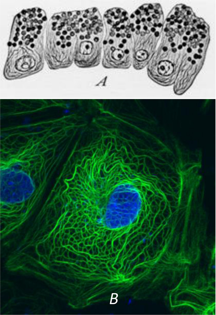
(A) Early observations of secretory epithelial cells in the mudpuppy pancreas. Note the “fibrillae” which most likely represent keratin IF bundles or tonofibrils. Work of Mathews in the laboratory of E.B. Wilson. From reference: (Wilson, 1928)). (B) A PtK2 epithelial cell fixed and processed for immunostaining with anti-keratin. Note the similarities in the tonofibrils seen in the preparations spanning 80 years of research.
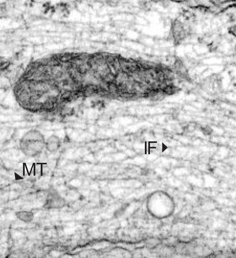
Electron micrograph of a human fibroblast showing the cytoplasm containing numerous 10 nm vimentin IF (IF) and a few microtubules (MT).
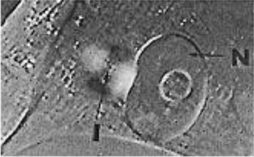
A living BHK-21 cell observed during the early stages of cell spreading by polarized light microscopy. The birefringent sphere comprised of dark and bright sectors surrounds an isotropic region (I). This structure is closely associated with the nucleus (N). From reference: (Goldman and Follett, 1970)
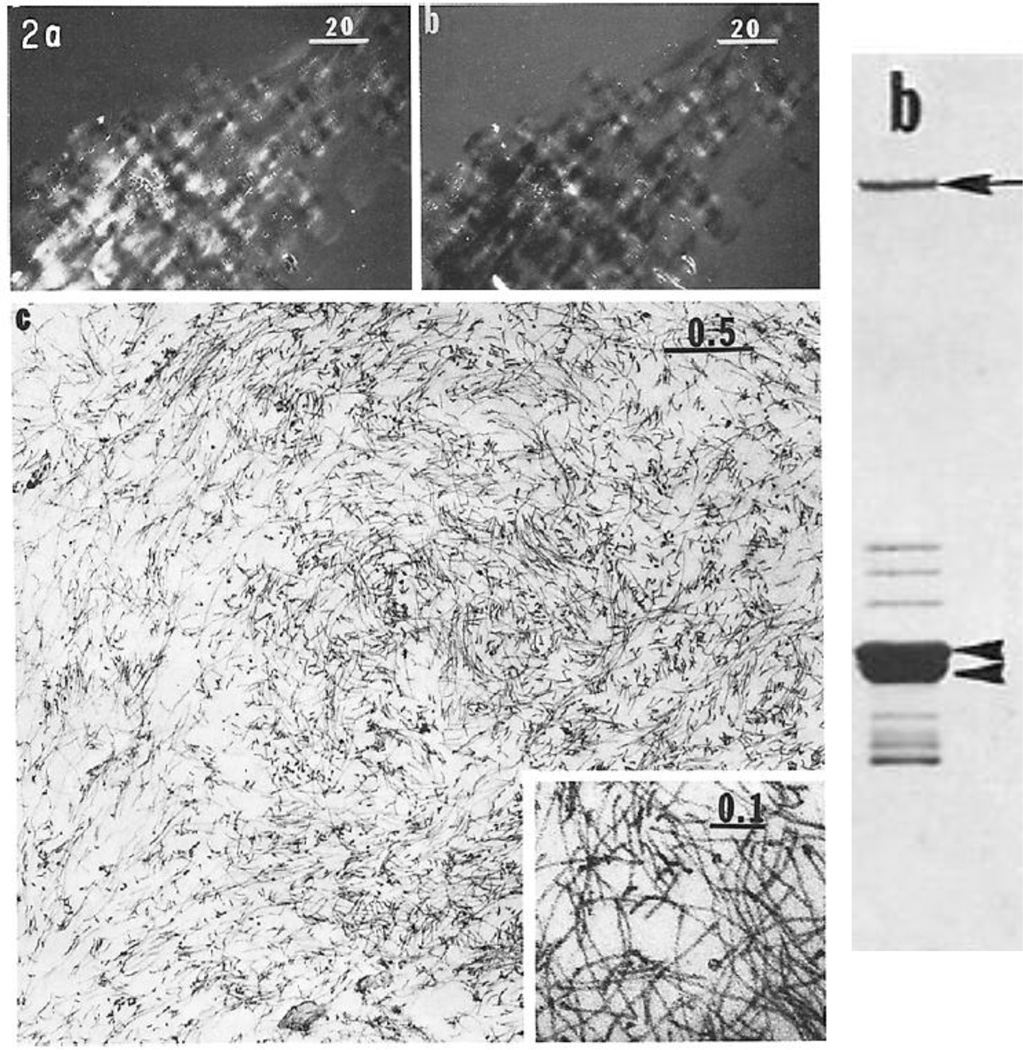
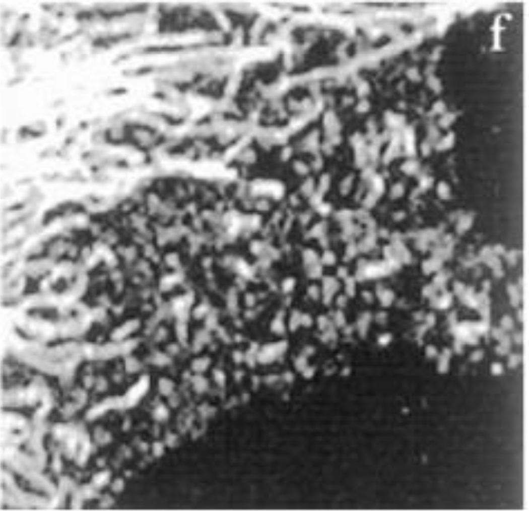
Left panel; Panels a and b show pellets of isolated birefringent juxtanuclear caps at opposite compensator settings; panel c is an electron micrograph of a thin section of a similar pellet showing large numbers of IF (from reference: (Starger and Goldman, 1977)). The right panel shows a Coomassie stained SDS-gel of intact IF obtained from the caps (from reference: (Starger et al., 1978)).
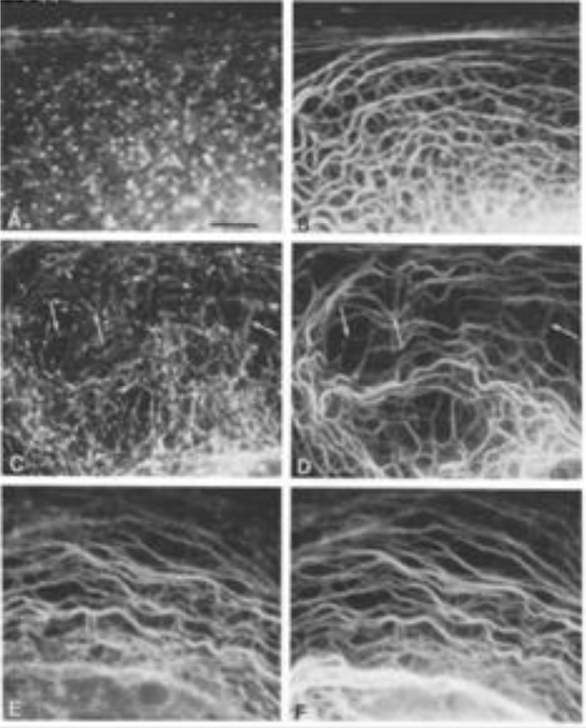
Purified biotinylated Type I keratin was microinjected into PtK2 cells and cells were fixed and processed for immunofluorescence with anti-biotin (Fig 5, A, C, E) and anti-keratin (Fig. 5 B, D, Figure legends F) at different time intervals (20 min [A,B], 1hr [C,D] and 4hr [E,F] post-microinjection). Taken from reference: (Miller et al., 1991).

A region of a BHK-21 cell expressing GFP-vimentin before (top) and immediately following photobleaching (00); 6 min after bleaching (06) and 18 min following bleaching (18)). Figure is from reference: (Yoon et al., 1998).
A BHK21 cell fixed and processed for immunofluorescence with anti-vimentin. Shown are different structural forms of IF including vimentin particles which give rise to squiggles or short IF and, through end domain interactions, form long IF. From reference: (Prahlad et al., 1998)
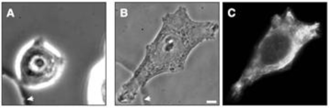
An MCF-7 cell prior to (A) and following the microinjection of soluble vimentin. The cell was fixed and processed for immunofluorescence with vimentin antibody at 5 hrs. A, B are phase contrast images and C is the same cell as B, but observed with fluorescence optics). From reference: (Mendez et al., 2010).
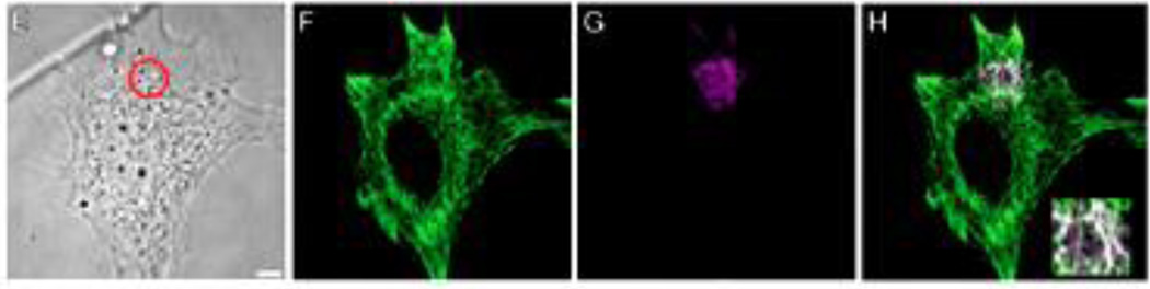
A mouse embryo fibroblast (mEF) expressing PA-Rac1 before (E) and fixed within 5 sec. after spot irradiation. Subsequently this cell was processed for double label immunofluorescence with vimentin (green) and vimentin pSer38 (magenta) antibodies. Insert shows that this latter antibody stains vimentin IF only in the irradiated spot). Taken from reference: (Helfand et al., 2011).
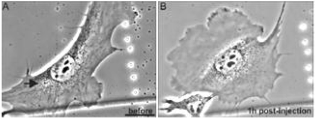
Serum deprived live mEF before (A) and at 1hr following microinjection of peptide 2B2. The arrow shows site of injection where a lamellipodium formed (upper left)). Taken from reference: (Helfand et al., 2011)
References
-
- Aebi U, Cohn J, Buhle L, Gerace L. The nuclear lamina is a meshwork of intermediate-type filaments. Nature. 1986;323:560–564. - PubMed
Publication types
MeSH terms
Substances
Grants and funding
LinkOut - more resources
Full Text Sources
Other Literature Sources
Miscellaneous
