Comparative Study
. 2012 May;55(5):1518-28.
doi: 10.1002/hep.25500. Epub 2012 Mar 18.

Toll-like receptor 4-dependent Kupffer cell activation and liver injury in a novel mouse model of parenteral nutrition and intestinal injury

Affiliations
Comparative Study

Toll-like receptor 4-dependent Kupffer cell activation and liver injury in a novel mouse model of parenteral nutrition and intestinal injury

Karim C El Kasmi et al. Hepatology. 2012 May.

Abstract

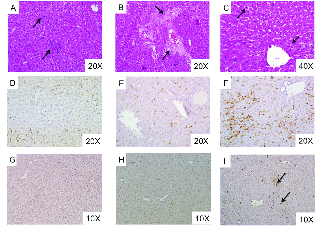
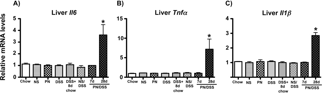
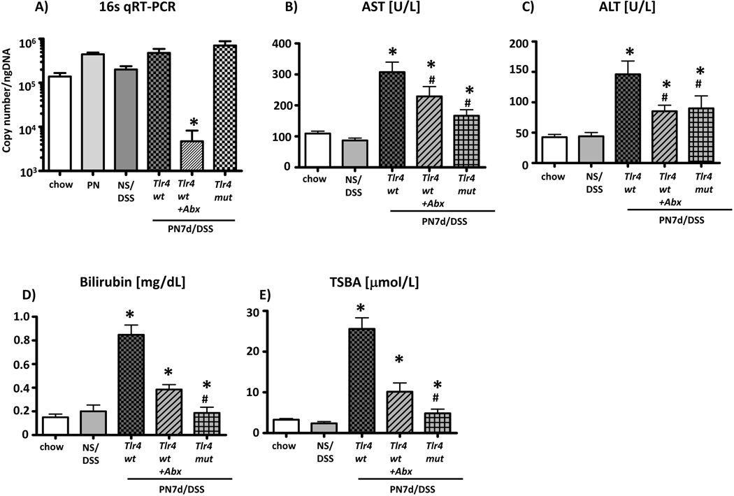
Infants with intestinal failure who are parenteral nutrition (PN)-dependent may develop cholestatic liver injury and cirrhosis (PN-associated liver injury: PNALI). The pathogenesis of PNALI remains incompletely understood. We hypothesized that intestinal injury with increased intestinal permeability combined with administration of PN promotes lipopolysaccharide (LPS)-Toll-like receptor 4 (TLR4) signaling dependent Kupffer cell (KC) activation as an early event in the pathogenesis of PNALI. We developed a mouse model in which intestinal injury and increased permeability were induced by oral treatment for 4 days with dextran sulphate sodium (DSS) followed by continuous infusion of soy lipid-based PN solution through a central venous catheter for 7 (PN7d/DSS) and 28 (PN28d/DSS) days. Purified KCs were probed for transcription of proinflammatory cytokines. PN7d/DSS mice showed increased intestinal permeability and elevated portal vein LPS levels, evidence of hepatocyte injury and cholestasis (serum aspartate aminotransferase, alanine aminotransferase, bile acids, total bilirubin), and increased KC expression of interleukin-6 (Il6), tumor necrosis factor α (Tnfα), and transforming growth factor β (Tgfβ). Markers of liver injury remained elevated in PN28d/DSS mice associated with lobular inflammation, hepatocyte apoptosis, peliosis, and KC hypertrophy and hyperplasia. PN infusion without DSS pretreatment or DSS pretreatment alone did not result in liver injury or KC activation, even though portal vein LPS levels were elevated. Suppression of the intestinal microbiota with broad spectrum antibiotics or ablation of TLR4 signaling in Tlr4 mutant mice resulted in significantly reduced KC activation and markedly attenuated liver injury in PN7d/DSS mice.

Conclusion: These data suggest that intestinal-derived LPS activates KC through TLR4 signaling in early stages of PNALI.


Conflict of interest statement

CONFLICT OF INTEREST.

The authors have no conflicts of interest to disclose.

Figures

Figure 1
Figure 1. Increased intestinal permeability combined with PN infusion in mice induces liver injury and cholestasis
(A) DSS pre-treated (n=10), PN/DSS7d (n=6), and untreated control mice (chow; n=5) were gavaged with FITC labeled-dextran to determine intestinal permeability. Untreated control mice not gavaged with FITC labeled-dextran are shown as a base line control (n=8). 4hrs later fluorescence was measured in portal serum specimens. Intestinal permeability was increased after DSS pre-treatment (DSS) and remained elevated after PN/DSS7d treatment. *p<0.05 DSS and PN/DSS7d vs. chow; determined by One-Way ANOVA. (B) Increased absorption of LPS in mice with increased intestinal permeability. Portal serum LPS levels [EU/L] were measured in untreated chow fed controls (n=8), DSS pre-treated (n=8), DSS+8d chow (n=8), and PN/DSS7d mice (n=6). DSS treatment increased LPS absorption, which was further significantly increased in PN/DSS mice compared to the other groups; *p<0.05 by One-Way ANOVA. (C–F) The combination of PN infusion with increased intestinal permeability induced hepatocyte injury (C,D) and cholestasis (E,F) in mice after 7 and 28 days of PN; *p<0.05 PN/DSS vs. controls by One-Way ANOVA. Mean values and standard error of mean are depicted.
Figure 2
Figure 2. PN/DSS treatment induces histological liver lesions
(A–C) H&E stained liver from PN/DSS28d mice shows focal areas of inflammation (arrows in panel A), loss of hepatocytes (arrows in panel B) and focal hepatocellular apoptosis (arrows in panel C pointing to Councilman bodies). (D–F) Immunohistochemistry using pan macrophage marker F4/80 demonstrates Kupffer cell hyperplasia and hypertrophy (arrow in panel F) in PN/DSS28d mice (F) as compared to PN/DSS7d mice (E) and chow fed control mice (D).
Figure 3
Figure 3. Kupffer cell activation in PN/DSS mice
(A–C) qRT-PCR analysis depicting relative transcript levels of IL6 (A), TNFα (B), and TGFβ (C) mRNA in purified Kupffer cells. Kupffer cells derived from LPS injected mice (LPS) served as a positive control for innate activation of Kupffer cells. Kupffer cell activation is increased in PN/DSS7d wild type mice (PN/DSS7d tlr4wt) compared to controls. Note that DSS treatment alone (DSS, DSS+8d chow, NS/DSS) or PN infusion in the absence of DSS pre-treatment (PN) does not induce Kupffer cell activation. Kupffer cell activation is attenuated after suppression of bowel flora (PN/DSS7d tlr4wt+Abx) or ablation of TLR4 signaling (PN/DSS7d tlr4mut) compared to Kupffer cells from PN/DSS7d wild type mice (PN/DSS7d tlr4wt). Data were normalized to Hypoxanthine-phosphoribosyl-transferase 1 (Hprt1) as endogenous control gene. mRNA levels are expressed relative to results obtained from untreated chow fed controls. Mean values and standard error of mean (SEM) of two independent Kupffer cell isolations that were analyzed in triplicate are depicted. *p<0.05 PN/DSS7d tlr4wt and LPS vs. controls; #p<0.05 PN/DSS7d tlr4wt+Abx and PN/DSS7d tlr4mut vs. PN/DSS7d tlr4wt; determined by One-Way ANOVA.
Figure 4
Figure 4. Inflammatory gene transcription is increased in livers from PN/DSS28d mice
qRT-PCR analysis depicting relative transcript levels of IL6 (A), TNFα (B), and IL1β (C) mRNA in whole liver homogenates. Elevated transcription was present at 28 but not 7 days of PN/DSS. Data were normalized to Hypoxanthine-phosphoribosyl-transferase 1 (Hprt1) as endogenous control gene. mRNA levels are expressed relative to results obtained from untreated chow fed controls. Mean values and SEM from n=5 samples tested in triplicate are depicted. *p<0.05 PN/DSS28d vs. all other groups; determined by One-Way ANOVA.
Figure 5
Figure 5. Liver injury is attenuated after suppression of bowel flora or ablation of TLR4 signaling
(A) qRT-PCR to determine copy number of bacterial 16S DNA genes in colonic stool samples after oral antibiotic treatment. Note significant reduction in bacterial load in stool from PN/DSS7d tlr4wt+Abx mice. Note that ablation of TLR4 signaling does not reduce bacterial load in stool of PN/DSS7d tlr4mut mice compared to wild type mice; *p<0.05 PN/DSS7d tlr4wt+Abx vs. other groups; determined by One-Way ANOVA. Hepatocyte injury (B-C) and cholestasis (D-E) were attenuated in PN/DSS wild type mice that were treated with oral antibiotics (PN/DSS7d tlr4 wt+Abx; n=11) or had non-functional TLR4 signaling (PN/DSS7d tlr4mut; n=12) compared to PN/DSS wild type mice (PN/DSS7d tlr4wt; n=15). NS/DSS mice (n=12) and untreated chw fed mice (n=20) served as controls. Mean values and SEM are depicted. *p<0.05 PN/DSS tlr4wt vs. controls and PN/DSS7d tlr4wt+Abx and PN/DSS7d tlr4mut vs. PN/DSS tlr4wt ; # p<0.05 PN/DSS7d tlr4wt+Abx and PN/DSS7d tlr4mut vs. controls; determined by One-Way ANOVA.

References

    1. Wilmore DW, Dudrick SJ. Growth and development of an infant receiving all nutrients exclusively by vein. JAMA. 1968;203:860–864. - PubMed
    1. Beale EF, Nelson RM, Bucciarelli RL, Donnelly WH, Eitzman DV. Intrahepatic cholestasis associated with parenteral nutrition in premature infants. Pediatrics. 1979;64:342–347. - PubMed
    1. Goulet O, Ruemmele F. Causes and management of intestinal failure in children. Gastroenterology. 2006;130:S16–S28. - PubMed
    1. Kaufman SS, Loseke CA, Lupo JV, Young RJ, Murray ND, Pinch LW, et al. Influence of bacterial overgrowth and intestinal inflammation on duration of parenteral nutrition in children with short bowel syndrome. J Pediatr. 1997;131:356–361. - PubMed
    1. Pierro A, van Saene HK, Donnell SC, Hughes J, Ewan C, Nunn AJ, et al. Microbial translocation in neonates and infants receiving long-term parenteral nutrition. Arch Surg. 1996;131:176–179. - PubMed

Publication types

MeSH terms