. 2011 Nov 27;480(7378):538-42.
doi: 10.1038/nature10664.

Response to self antigen imprints regulatory memory in tissues

Affiliations

Response to self antigen imprints regulatory memory in tissues

Michael D Rosenblum et al. Nature. .

Abstract

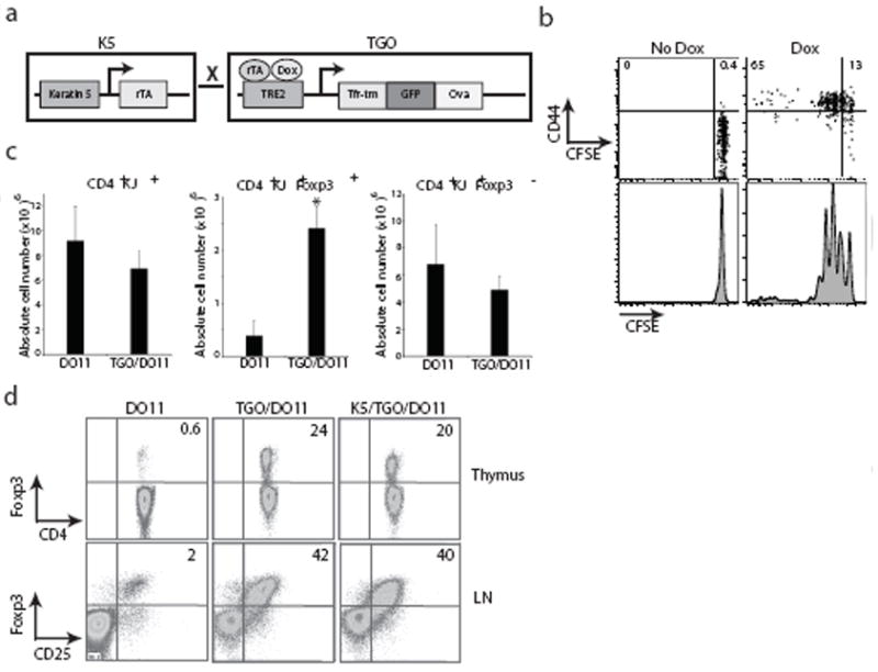
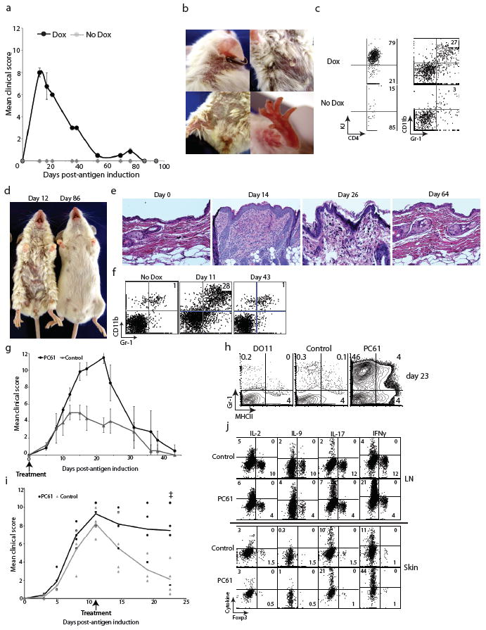
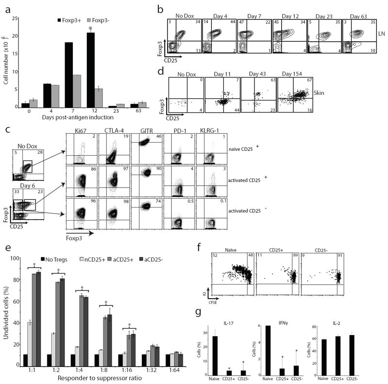
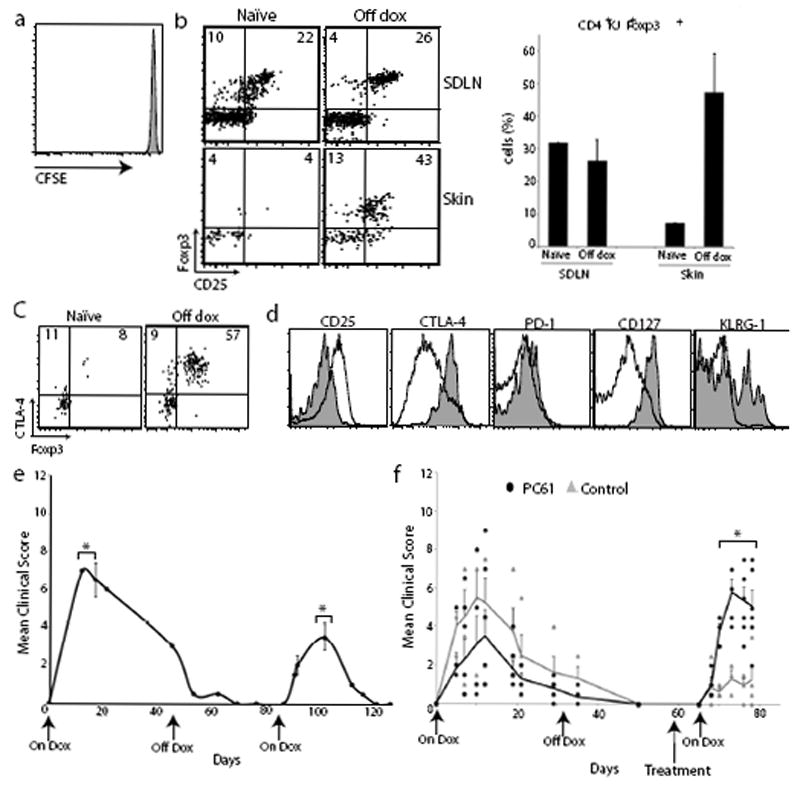
Immune homeostasis in tissues is achieved through a delicate balance between pathogenic T-cell responses directed at tissue-specific antigens and the ability of the tissue to inhibit these responses. The mechanisms by which tissues and the immune system communicate to establish and maintain immune homeostasis are currently unknown. Clinical evidence suggests that chronic or repeated exposure to self antigen within tissues leads to an attenuation of pathological autoimmune responses, possibly as a means to mitigate inflammatory damage and preserve function. Many human organ-specific autoimmune diseases are characterized by the initial presentation of the disease being the most severe, with subsequent flares being of lesser severity and duration. In fact, these diseases often spontaneously resolve, despite persistent tissue autoantigen expression. In the practice of antigen-specific immunotherapy, allergens or self antigens are repeatedly injected in the skin, with a diminution of the inflammatory response occurring after each successive exposure. Although these findings indicate that tissues acquire the ability to attenuate autoimmune reactions upon repeated responses to antigens, the mechanism by which this occurs is unknown. Here we show that upon expression of self antigen in a peripheral tissue, thymus-derived regulatory T cells (T(reg) cells) become activated, proliferate and differentiate into more potent suppressors, which mediate resolution of organ-specific autoimmunity in mice. After resolution of the inflammatory response, activated T(reg) cells are maintained in the target tissue and are primed to attenuate subsequent autoimmune reactions when antigen is re-expressed. Thus, T(reg) cells function to confer 'regulatory memory' to the target tissue. These findings provide a framework for understanding how T(reg) cells respond when exposed to self antigen in peripheral tissues and offer mechanistic insight into how tissues regulate autoimmunity.


Figures

Figure 1
Figure 1. Characterization of K5/TGO/DO11 mice
(a) Construct for double transgenic mice expressing ovalbumin (Ova) driven by the cytokeratin-5 (K5) promoter in a tetracycline-inducible fashion. (b) Lymph node cells from DO11 TCR-transgenic mice were labeled with CFSE and injected into K5/TGO mice. Recipient mice were fed doxycycline chow and DO11 cell proliferation (CFSE dilution) and CD44 expression was measured 3 days later by flow cytometry. (c) SDLN cell numbers from DO11 and TGO/DO11 mice in the absence of doxycycline treatment. (d) Thymus and SDLN cells from DO11, TGO/DO11, and K5/TGO/DO11 mice in the absence of doxycycline treatment. Thymocytes are gated on CD4+KJ+CD8- cells. Lymph node cells are gated on CD4+KJ+ cells. *P < 0.05 (t-test). Error bars represent s.d. Results are representative of 3 replicate experiments with n=3-4 mice/group.
Figure 2
Figure 2. K5/TGO/DO11 mice develop autoimmune skin disease that resolves spontaneously
(a) Mean clinical scores of K5/TGO/DO11 mice left untreated or fed doxycycline chow. (b) Skin lesions in a representative K5/TGO/DO11 mouse at the height of clinical disease. (c) Flow cytometry of skin-infiltrating cells in K5/TGO/DO11 mice treated with doxycycline for 12 days. (d) Representative mice from the height of disease (Day 12) and after disease resolution (Day 86). (e) Skin histology of K5/TGO/DO11 mice after beginning doxycycline. (f) Flow cytometry of skin-infiltrating cells in K5/TGO/DO11 mice at 11 and 43 days after beginning doxycycline. (g) Disease scores of K5/TGO/DO11 mice treated with anti-CD25 monoclonal antibody (PC61) or control (PBS or isotype control antibody) at 10 days and 3 days prior to beginning doxycycline. (h) Flow cytometry of skin-infiltrating cells in K5/TGO/DO11 mice untreated or treated with PC61 at 23 days after beginning doxycycline. (i) Disease scores of K5/TGO/DO11 mice treated with PC61 or isotype control antibody at the height of disease. Results are pooled data from two replicate experiments with n=3-4 mice/group. (j) Intracellular cytokine stains of SDLN cells and skin-infiltrating cells from PC61- or isotype control-treated (prior to antigen induction) K5/TGO/DO11 mice at 11 days after beginning doxycycline. Gated on CD4+KJ+ cells. Error bars represent s.d. Results are representative of 3 replicate experiments with n=3-4 mice/group. Ŧ denotes euthanization of two PC61-treated mice secondary to non-resolving skin disease.
Figure 3
Figure 3. Treg cells are activated upon induction of peripheral antigen
(a) Quantification of CD4+KJ+Foxp3+ and CD4+KJ+Foxp3- cell numbers from SDLNs of K5/TGO/DO11 mice after beginning doxycycline. (b) Flow cytometry of SDLN cells from K5/TGO/DO11 mice after beginning doxycycline. Gated on CD4+KJ+ cells. (c) Flow cytometry of SDLN cells from K5/TGO/DO11 mice left untreated (naïve CD25+) or treated for 6 days with doxycycline (activated CD25+ or CD25- populations). (d) Flow cytometry of skin-infiltrating cells from K5/TGO/DO11 mice after beginning doxycycline. Gated on live CD4+KJ+ cells. (e) In vitro suppression assay using sorted CD4+KJ+Foxp3+CD25+ cells from non-induced naïve mice (nCD25+), CD4+KJ+Foxp3+CD25+ cells from doxycycline-treated mice (activated CD25+, aCD25+), or CD4+KJ+Foxp3+CD25- cells from doxycycline-treated mice (aCD25-) as suppressors and CFSE-labeled DO11 LN cells as responders. (f) In vivo suppression assay using suppressors (as in part e) mixed 1:1 with naïve CFSE-labeled Thy1.1+ DO11 LN cells and adoptively transferred into K5/TGO mice. Recipient mice were treated with doxycycline and DO11 cell proliferation and intracellular cytokine expression (g) were assayed 3 days later. Gated on Thy1.1+CD4+KJ+ cells. *P < 0.05 (t-test). Error bars represent s.d. Results are representative of 3 replicate experiments with n=3-4 mice/group except for in vitro and in vivo suppression assays, which are representative of 2 replicate experiments with n=2-3 mice/group.
Figure 4
Figure 4. Memory Treg cells attenuate skin disease upon re-expression of tissue antigen
(a) Flow cytometry of CFSE-labeled Thy1.1+ DO11+ SDLN cells 3 days after adoptive transfer into K5/TGO/DO11 mice that had been on doxycycline for >30 days and off for 20 days. (b) Flow cytometry and Treg cell percentages of SDLN and skin-infiltrating CD4+DO11 cells isolated from K5/TGO/DO11 mice that were on doxycycline for >30 days and off for >30 days. Gated on CD4+KJ+ cells. (c) Flow cytometry of CD4+DO11 cells from the skin of K5/TGO/DO11 mice that have not been treated with doxycycline (naïve) or on for >30 days and off doxycycline for >30 days. (d) Phenotype of CD4+Foxp3+ DO11 cells from skin (shaded) and SDLN (unshaded) from K5/TGO/DO11 mice that were on doxycycline for >30 days and off for >40 days. (e) Clinical skin disease upon re-starting doxycycline in K5/TGO/DO11 mice that had been on doxycycline for >30 days and off for >30 days. (f) Clinical skin disease of K5/TGO/DO11 mice treated with either PC61 or isotype control antibody prior to restarting doxycycline. In all mice, antigen was induced, skin disease developed, and disease had resolved before antibody treatment. Error bars represent standard error of samples within each group. Mean clinical scores from individual mice are shown. Results are representative of 3 replicate experiments with 3-4 mice/group except for (f) which is combined data from 2 replicate experiments with 2-4 mice/group. *P < 0.05 (t-test).

Comment in

References

    1. James WD. Andrews’ Diseases of the Skin: Clinical Dermatology. Saunders Elsevier; Philadelphia: 2006.
    1. Lara-Corrales I, Pope E. Autoimmune blistering diseases in children. Semin Cutan Med Surg. 2010;29:85–91. - PubMed
    1. Sabatos-Peyton CA, Verhagen J, Wraith DC. Antigen-specific immunotherapy of autoimmune and allergic diseases. Curr Opin Immunol. 2010;22:609–615. - PMC - PubMed
    1. Diamond I, Owolabi T, Marco M, Lam C, Glick A. Conditional gene expression in the epidermis of transgenic mice using the tetracycline-regulated transactivators tTA and rTA linked to the keratin 5 promoter. J Invest Dermatol. 2000;115:788–794. - PubMed
    1. Murphy KM, Heimberger AB, Loh DY. Induction by antigen of intrathymic apoptosis of CD4+CD8+TCRlo thymocytes in vivo. Science. 1990;250:1720–1723. - PubMed

Publication types