. 2011 Nov;6(6):809-822.
doi: 10.2217/fnl.11.45.

The role of neuregulin-1 in the response to nerve injury

Affiliations

The role of neuregulin-1 in the response to nerve injury

Florence R Fricker et al. Future Neurol. 2011 Nov.

Abstract

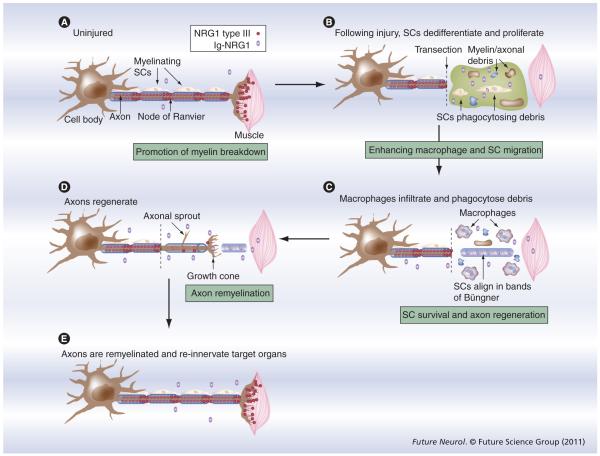
Axons and Schwann cells exist in a highly interdependent relationship: damage to one cell type invariably leads to pathophysiological changes in the other. Greater understanding of communication between these cell types will not only give insight into peripheral nerve development, but also the reaction to and recovery from peripheral nerve injury. The type III isoform of neuregulin-1 (NRG1) has emerged as a key signaling factor that is expressed on axons and, through binding to erbB2/3 receptors on Schwann cells, regulates multiple phases of their development. In adulthood, NRG1 is dispensable for the maintenance of the myelin sheath; however, this factor is required for both axon regeneration and remyelination following nerve injury. The outcome of NRG1 signaling depends on interactions with other pathways within Schwann cells such as Notch, integrin and cAMP signaling. In certain circumstances, this signaling pathway may be maladaptive; for instance, direct binding of Mycobacterium leprae onto erbB2 receptors produces excessive activation and can actually promote demyelination. Attempts to modulate this pathway in order to promote nerve repair will therefore need to give consideration to the exact isoform used, as well as how it is processed and the context in which it is presented to the Schwann cell.


Figures

Figure 1
Figure 1. Neuregulin-1 isoforms and signaling in the PNS
(A) The NRG1 isoform types expressed in the PNS. Isoforms are categorized into types depending on the composition of their N-terminal domains. Types I and II exist as pro-NRG1 forms in the membrane before being cleaved and shed, and type IIβ3 is directly secreted. All of these types signal in a paracrine fashion. Type III isoforms undergo a cleavage event but have a second TM domain that tethers them to the membrane, and thus type III signals in a juxtacrine fashion. TACE, BACE, ADAM10 and ADAM19 are all known to be capable of cleaving NRG1 isoforms. NRG1 isoform types IV, V and VI are also known to be expressed by humans and rodents; however, their function and expression patterns are as yet unknown in the PNS and consequently they have not been included in this figure [18]. Types I and II contain an Ig domain and are consequently referred to as Ig-NRG1s. Types IV and V also contain an Ig domain; however, due to their unknown role in the PNS, for the purpose of this article, Ig-NRG1 refers to type I and II isoforms. (B) The signaling pathways and transcription factors known to be modulated by NRG1–erbB2/3 signaling between axons and Schwann cells. NRG1 binds erbB receptors, resulting in heterodimerization and phosphorylation of the tyrosine residues present in the intracellular domains, creating docking sites for adaptor proteins, resulting in the induction of various signaling pathways, including PI3K–Akt, ERK1/2, FAK, Rac1 and cdc42, and the activation of calcineurin signaling pathways. NRG1 signaling is also known to regulate the expression of a nuclear variant of the erbB3 receptor (nuc-erbB3). These signaling pathways are known to regulate the activity of a range of transcription factors including NF-κB, NFAT, Yy1, Oct6 and Krox 20. These pathways modulate the Schwann cell responses to NRG1 signaling that include migration, survival, proliferation, differentiation and myelination. ADAM: A disintegrin and metalloprotease; BACE: β-secretase β-site of amyloid precursor protein-cleaving enzyme; Ig: Immunoglobulin; NRG: Neuregulin; TACE: TNF-α-converting enzyme; TM: Transmembrane.
Figure 2
Figure 2. Neuregulin-1 is required for axoglial signaling between axons and both myelinating Schwann cells and nonmyelinating Schwann cells
Electron micrographs of transverse sections of sural nerve from control mice (A) and mice in which neuregulin-1 (NRG1) is ablated in sensory axons during development using a Nav1.8 promoter-driven Cre recombinase (B). In the absence of NRG1, Remak bundles (arrow heads) are abnormally large, axons are not all separated by Schwann cell processes forming polyaxonal pockets and large diameter axons are present within the Remak bundle Δ (B). Such abnormal Remak bundles illustrate the importance of NRG1 in signaling between nonmyelinating Schwann cells and axons. There is also a small proportion of thinly myelinated axons (that express Nav1.8-Cre) in which NRG1 is ablated; the arrow identifies an axon that would usually be myelinated but is completely amyelinated. However, this axon is invested in Schwann cell cytoplasm and a basement membrane surrounds the fiber (B). Scale bars: 500 nm. Reproduced with permission from [56].
Figure 3
Figure 3. Neuregulin-1 is essential for remyelination following peripheral nerve injury
Electron micrographs of transverse sections of the sciatic nerve 8 weeks post-sciatic nerve crush injury in SLICK-A Cre;NRG1f/f mice in which neuregulin-1 (NRG1) is inducibly ablated in adulthood in a small percentage of axons that are labeled with yellow fluorescent protein (YFP) (B & C) and control (A). YFP-expressing axons are identified using immunoelectron microscopy and the resulting presence of a dark reaction product (asterisks). Control animals demonstrated effective remyelination of YFP-expressing axons (A). By contrast, YFP-positive axons from conditional NRG1-mutant (tamoxifen-treated SLICK-A Cre;NRG1fl/fl) mice either had a significantly thinner myelin sheath (B) or completely failed to elaborate a myelin sheath (C). Note that in (B), a neighboring axon that is YFP negative is normally myelinated, highlighting the importance of juxtacrine NRG1 signaling in remyelination following peripheral nerve injury. Scale bars: 500 nm. Reproduced with permission from [88].
Figure 4
Figure 4. Macrophages express erbB2, erbB3 and erbB4 and neuregulin-1 enhances macrophages motility
Peritoneal macrophages were cultured, these cells are identified by IBA1 immunostaining and nuclear staining with DAPI (A). These cells expressed erbB2, erbB3 and erbB4 (185-kDa band in western blot) (B) and showed enhanced motility in response to NRG1 assessed in the Boyden chamber assay (C). Error bars represent ± standard error of the mean (three independent experiments). The statistical tests used were one-way analysis of variance with Bonferroni post hoc ana lysis. Scale bar: 20 μm. *p < 0.05, **p < 0.001. CON: Control; NRG1: Neuregulin-1. Reproduced with permission from [98].
Figure 5
Figure 5. Neuregulin-1 is potentially involved at multiple stages of the nerve repair process following injury
Events after a peripheral nerve injury and the potential roles NRG1 may play in this process (dark boxes) (A–E). Following peripheral nerve injury – specifically a transection injury – a series of regulated events occur termed Wallerian degeneration. (B) Within 2 days following injury, the distal stump fragments and SCs begin clearance of axonal and myelin debris, and at the same time SCs proliferate and dedifferentiate. NRG1 has been implicated in promotion of this initial myelin clearance [80]. During the acute phase following injury, the expression levels of Ig-NRG1s, erbB2 and erbB3 increase in SCs, and this expression is sustained long after myelin clearance. Neuronal expression of NRG1 type III decreases and does not return to normal levels until axons re-innervate their target organs. (C) Macrophages infiltrate into the distal stump, and within 2 weeks, both macrophages and SCs complete the clearance of debris, and at the same time, dedifferentiated SCs align themselves in bands of Büngner. NRG1 is known to enhance macrophage motility, so it may play a role in macrophage recruitment following nerve injury, although this has not yet been shown in vivo [98]. (D) Axons then regenerate out of the proximal stump following SC tubes and remake contact with SCs. NRG1 is known to play a role in axon regeneration [88] and may also have a role in SC survival, particularly in chronically denervated nerve stumps. (E) Axons re-innervate their target organs and are remyelinated by SCs in order to regain full function. NRG1 is known to be essential for remyelination of axons following nerve injury [88]. NRG1 may therefore be involved at multiple stages of peripheral nerve repair. Ig: Immunoglobulin; NRG1: Neuregulin-1; SC: Schwann cell.

Similar articles

Cited by

References

    1. Martyn CN, Hughes RA. Epidemiology of peripheral neuropathy. J. Neurol. Neurosurg. Psychiatry. 1997;62(4):310–318. - PMC - PubMed
    1. Mohanna PN, Young RC, Wiberg M, Terenghi G. A composite poly-hydroxybutyrate-glial growth factor conduit for long nerve gap repairs. J. Anat. 2003;203(6):553–565. - PMC - PubMed
    1. Filbin MT. Myelin-associated inhibitors of axonal regeneration in the adult mammalian CNS. Nat. Rev. Neurosci. 2003;4(9):703–713. - PubMed
    1. Matsuyama T, Mackay M, Midha R. Peripheral nerve repair and grafting techniques: a review. Neurol. Med. Chir. 2000;40(4):187–199. - PubMed
    1. Webber C, Zochodne D. The nerve regenerative microenvironment: early behavior and partnership of axons and Schwann cells. Exp. Neurol. 2010;223(1):51–59. - PubMed

LinkOut - more resources