A rare case of subependymoma with an atypical presentation: a case report
- PMID: 22121350
- PMCID: PMC3223030
- DOI: 10.1159/000333061
A rare case of subependymoma with an atypical presentation: a case report
Abstract
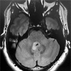
A rare case of subependymoma in a young patient presenting with sensory dysesthesia is reported. Computed tomography scan and magnetic resonance imaging revealed a posterior fossa mass occluding the fourth ventricle with infiltration to the right side immediately behind the pontine tegmentum and impinging on the right spinothalamic tract. Postoperative tumor histopathology revealed the classical appearance of subependymoma. Subependymoma is a rare, asymptomatic, slow-growing, low-grade glioma of the central nervous system. If symptomatic, the clinical features are commonly secondary to hydrocephalus, but subependymoma presenting with sensory dysesthesia has never been reported in the literature.
Keywords: Intraventricular tumor; Sensory dysesthesia; Subependymoma.
Figures


Similar articles
-
Symptomatic subependymoma--a case report.J Korean Med Sci. 1990 Jun;5(2):111-5. doi: 10.3346/jkms.1990.5.2.111. J Korean Med Sci. 1990. PMID: 2278665 Free PMC article.
-
Recurrent subependymoma of fourth ventricle with unusual atypical histological features: A case report.Pathol Int. 2015 Aug;65(8):438-42. doi: 10.1111/pin.12316. Epub 2015 Jun 8. Pathol Int. 2015. PMID: 26059172
-
Subependymoma at the foramen of Monro presenting with intermittent hydrocephalus: case report and review of the literature.J La State Med Soc. 2010 Jul-Aug;162(4):214-7. J La State Med Soc. 2010. PMID: 20882814
-
Subependymoma in the third ventricle in a child.Clin Imaging. 2004 Sep-Oct;28(5):381-4. doi: 10.1016/S0899-7071(03)00203-1. Clin Imaging. 2004. PMID: 15471674 Review.
-
Massive symptomatic subependymoma of the lateral ventricles: case report and review of the literature.Neuroradiology. 2005 Mar;47(3):183-8. doi: 10.1007/s00234-005-1342-3. Epub 2005 Feb 9. Neuroradiology. 2005. PMID: 15702322 Review.
Cited by
-
Intraventricular Subependymoma With Obstructive Hydrocephalus: A Case Report and Literature Review.Cureus. 2024 Jan 19;16(1):e52563. doi: 10.7759/cureus.52563. eCollection 2024 Jan. Cureus. 2024. PMID: 38371163 Free PMC article.
-
Pedunculated intraventricular subependymoma: Review of the literature and illustration of classical presentation through a clinical case.Surg Neurol Int. 2014 Jul 30;5:117. doi: 10.4103/2152-7806.137837. eCollection 2014. Surg Neurol Int. 2014. PMID: 25101212 Free PMC article.
-
Lateral Horizontal Head Position Approach for the Lateral and Anterior Third Ventricles: A Subependymoma Clinical Case and Literature Review.Asian J Neurosurg. 2022 Oct 18;17(4):642-646. doi: 10.1055/s-0042-1757724. eCollection 2022 Dec. Asian J Neurosurg. 2022. PMID: 36570767 Free PMC article.
-
A First Case Report of Subependymoma in PTPN11 Mutation-Associated Noonan Syndrome.Case Rep Neurol Med. 2019 Sep 16;2019:6091059. doi: 10.1155/2019/6091059. eCollection 2019. Case Rep Neurol Med. 2019. PMID: 31637070 Free PMC article.
-
Atypical hemorrhagic presentation of a fourth ventricle subependymoma: case report.Neurol Med Chir (Tokyo). 2013;53(11):828-31. doi: 10.2176/nmc.cr2012-0292. Epub 2013 Oct 21. Neurol Med Chir (Tokyo). 2013. PMID: 24140775 Free PMC article. Review.
References
-
- Matsumara A, Ahyai A, Hori A, Schaake T. Intracerebral subependymomas: clinical and neuropathological analyses with special reference to the possible existence of a less benign variant. Acta Neurochir. 1989;96:15–25. - PubMed
-
- Prayson RA, Suh JH. Subependymomas: clinicopathologic study of 14 tumors, including comparative MIB-1 immunohistochemical analysis with other ependymal neoplasms. Arch Pathol Lab Med. 1999;123:306–309. - PubMed
-
- Scheinker I. Subependymoma: a newly recognized tumor of subependymal derivation. J Neurosurg. 1945;2:232–240.
-
- Koeller KK, Sandberg GD. Cerebral intraventricular neoplasms: radiologic-pathologic correlation. Radiographics. 2002;22:1473–1505. - PubMed
-
- Scheithauer B. Symptomatic subependymoma: report of 21 cases with review of the literature. J Neurosurg. 1978;49:689–696. - PubMed
Publication types
LinkOut - more resources
Full Text Sources

