. 2012:2012:517529.
doi: 10.1155/2012/517529. Epub 2011 Nov 9.

Hyphal growth in human fungal pathogens and its role in virulence

Affiliations

Hyphal growth in human fungal pathogens and its role in virulence

Alexandra Brand. Int J Microbiol. 2012.

Abstract

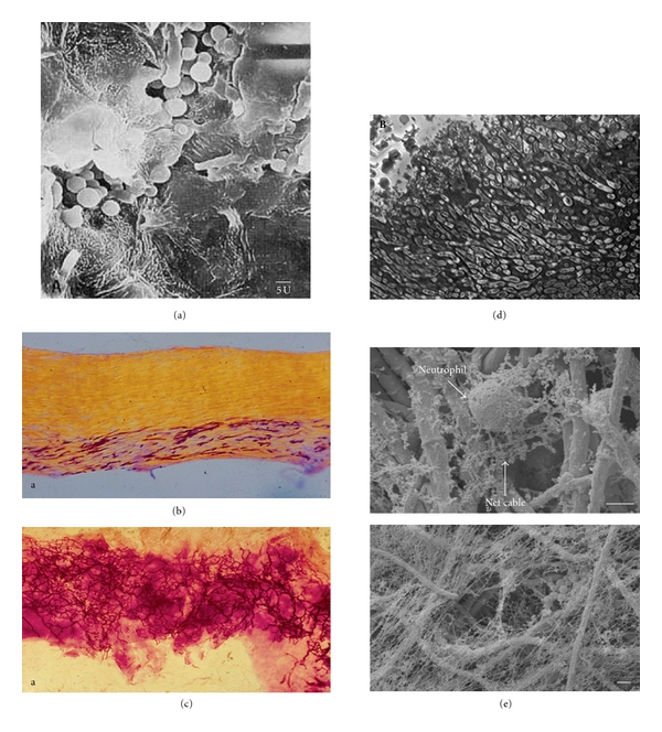
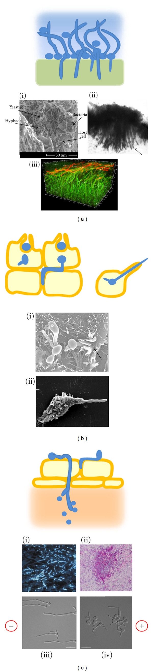
Most of the fungal species that infect humans can grow in more than one morphological form but only a subset of pathogens produce filamentous hyphae during the infection process. This subset is phylogenetically unrelated and includes the commonly carried yeasts, Candida albicans, C. dubliniensis, and Malassezia spp., and the acquired pathogens, Aspergillus fumigatus and dermatophytes such as Trichophyton rubrum and T. mentagrophytes. The primary function of hypha formation in these opportunistic pathogens is to invade the substrate they are adhered to, whether biotic or abiotic, but other functions include the directional translocation between host environments, consolidation of the colony, nutrient acquisition and the formation of 3-dimensional matrices. To support these functions, polarised hyphal growth is co-regulated with other factors that are essential for normal hypha function in vivo.


Figures

Figure 1
Figure 1
In vivo growth of filamentous fungal pathogens. (a) Hyphae of Malassezia globosa translocate deeper within the keratinised epidermal layer where they establish new colonies and revert to growth as yeast [13]. (b) Dermatophyte hyphae follow the keratinised layers that run parallel to the nail surface [14]. (c) Hyphae of Candida albicans growing in a multidirectional manner within the nail, often forming helical twists [14]. (d) Histological section of aspergilloma in the lung showing the tightly packed Aspergillus fumigatus hyphae surrounded by a matrix material and with no immune-cell infiltrate [15]. (e) Production of neutrophil extracellular traps (NETs) against A. fumigatus hyphae where, unlike the safe haven of a fungus-derived biofilm matrix, hyphae are instead imprisoned in a host-derived matrix of neutrophil DNA and calprotectin, a protein which chelates the divalent cations that are required for fungal growth [16, 17].
Figure 2
Figure 2
Models and examples of hyphal invasion in vivo. (a) Biofilms form on mucosal and abiotic surfaces by initial adhesion of yeast cells, followed by hypha germination and the deposition of extracellular matrix polysaccharides (blue). On mucosa or soft silicones, hyphae penetrate the underlying layers. (i) Rat denture biofilm formation after 48 h [37]. (ii) Hyphae penetrate the silicone of a voice prosthesis, causing it to expand and stiffen [7]. (iii) In silico construction of a biofilm showing the thick hyphal matrix (green) and leading edge of β-glucan deposition at the hyphal tips (red) [38]. (b) Induced uptake of yeast and newly germinating hyphae by epithelial cells or phagocytosis by macrophages is followed by sustained polarised growth, which breaches the host cell plasma membrane and permits the escape of the fungus. (i) C. albicans hyphae are engulfed by epithelial cells during induced uptake [39]. (ii) C. albicans avoids being killed by a macrophage by undergoing morphogenesis and breaching the macrophage membrane [40]. (c) Active penetration of endothelial cells and reversion to yeast growth in the tissue below. (i) Biopsy of murine lung with invasive aspergillosis, showing septate hyphae stained with blancophor [41]. (ii) Histological section of murine kidney showing C. albicans lesion containing yeast, hyphae, and infiltrate of neutrophils (courtesy D. MacCallum). (iii) Galvanotropism: C. albicans wild-type hyphae orient towards the cathode when grown in an applied electric field (10 V/cm) [42]. (iv) All tropic responses were abolished in the rsr1Δ mutant, which was attenuated in virulence. Cell polarity was maintained, but hyphal tip directionality was erratic [42].

References

    1. Benham RW. The cultural characteristics of Pityrosporum ovale; a lipophilic fungus. Journal of Investigivative Dermatology. 1939;2:187–203.
    1. Xu J, Saunders CW, Hu P, et al. Dandruff-associated Malassezia genomes reveal convergent and divergent virulence traits shared with plant and human fungal pathogens. Proceedings of the National Academy of Sciences of the United States of America. 2007;104(47):18730–18735. - PMC - PubMed
    1. Ro BI, Dawson TL. The role of sebaceous gland activity and scalp microfloral metabolism in the etiology of seborrheic dermatitis and dandruff. Journal of Investigative Dermatology Symposium Proceedings. 2005;10(3):194–197. - PubMed
    1. Mccormick A, Loeffler J, Ebel F. Aspergillus fumigatus: contours of an opportunistic human pathogen. Cellular Microbiology. 2010;12(11):1535–1543. - PubMed
    1. Kriengkauykiat J, Ito JI, Dadwal SS. Epidemiology and treatment approaches in management of invasive fungal infections. Clinical Epidemiology. 2011;3(1):175–191. - PMC - PubMed

LinkOut - more resources