Review
. 2011 Dec;125(6):797-824.
doi: 10.1037/a0026187.

Memory modulation

Affiliations
Review

Memory modulation

Benno Roozendaal et al. Behav Neurosci. 2011 Dec.

Abstract

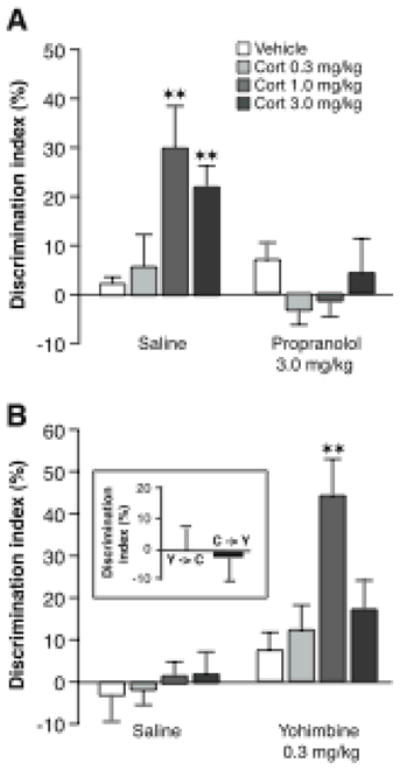
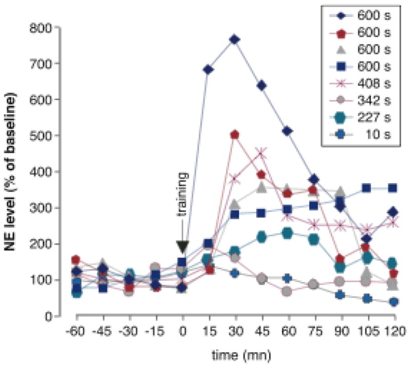
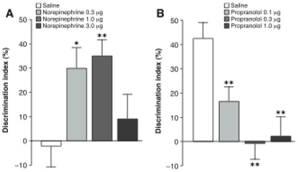
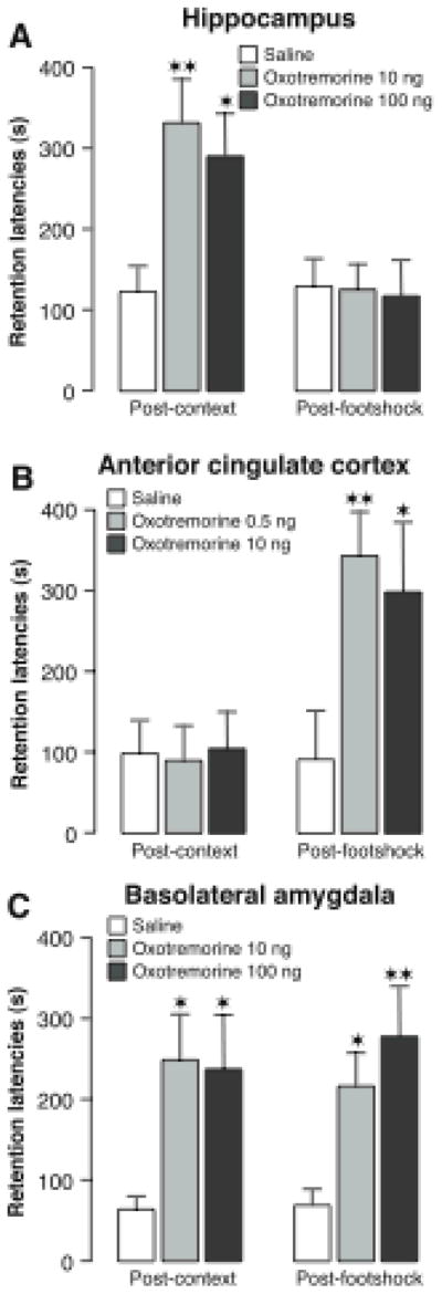
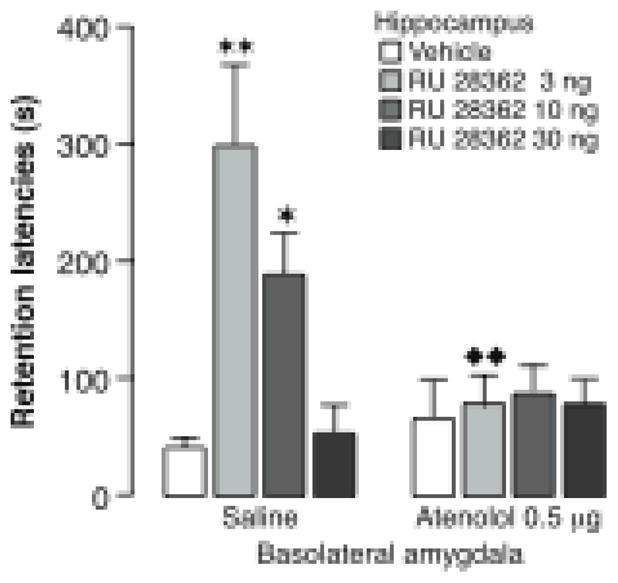
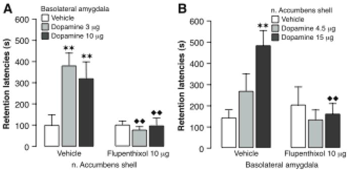
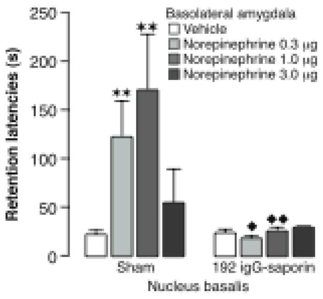
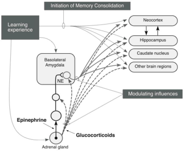
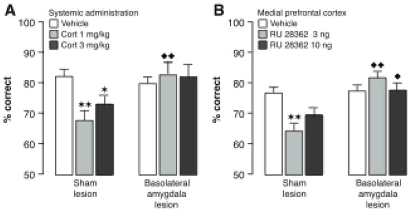
Our memories are not all created equally strong: Some experiences are well remembered while others are remembered poorly, if at all. Research on memory modulation investigates the neurobiological processes and systems that contribute to such differences in the strength of our memories. Extensive evidence from both animal and human research indicates that emotionally significant experiences activate hormonal and brain systems that regulate the consolidation of newly acquired memories. These effects are integrated through noradrenergic activation of the basolateral amygdala that regulates memory consolidation via interactions with many other brain regions involved in consolidating memories of recent experiences. Modulatory systems not only influence neurobiological processes underlying the consolidation of new information, but also affect other mnemonic processes, including memory extinction, memory recall, and working memory. In contrast to their enhancing effects on consolidation, adrenal stress hormones impair memory retrieval and working memory. Such effects, as with memory consolidation, require noradrenergic activation of the basolateral amygdala and interactions with other brain regions.


Figures

Figure 1
Figure 1
Glucocorticoid effects on memory consolidation for object recognition training require noradrenergic activation. A, Immediate posttraining administration of the β-adrenoceptor antagonist propranolol (3.0 mg/kg, sc) blocked the corticosterone-induced enhancement of object recognition memory in naïve rats. B, The α2-adrenoceptor antagonist yohimbine (0.3 mg/kg, sc) enabled corticosterone effect on object recognition memory in habituated rats. Inset, Posttraining injections of yohimbine (0.3 mg/kg, sc) and corticosterone (1.0 mg/kg, sc) separated by a 4-hour delay did not induce memory enhancement. Y→C; Yohimbine administered immediately after training and corticosterone 4 hours later; C→Y; corticosterone administered immediately after training and yohimbine 4 hours later. Results represent discrimination index (mean ± SEM) in percentage on a 24-hour retention trial. The discrimination index was calculated as the difference in time spent exploring the two objects, expressed as the ratio of the total time spent exploring both objects. ★ ★, P < 0.01 as compared to the corresponding vehicle group. From Roozendaal et al., 2006a.
Figure 2
Figure 2
Norepinephrine levels in the amygdala in individual animals following inhibitory avoidance training. Percent of baseline norepinephrine following inhibitory avoidance training is graphed for each individual rat. The key notes retention score on the following day. Amygdala norepinephrine levels correlate with 24-hour retention performance. Correlation values for the first five posttraining samples varied from +0.75 to +0.92. From McIntyre et al., 2002.
Figure 3
Figure 3
Noradrenergic activation of the basolateral amygdala modulates consolidation of object recognition memory. A, Enhancing effects of posttraining intra-basolateral amygdala infusions of norepinephrine on 24-hour object recognition memory. Saline-infused controls displayed no evidence of memory of 3 minutes of training. The retention performance of groups given norepinephrine (0.3 or 1.0 μg in 0.2 μl) after training was significantly better than that of saline controls. Data presented as discrimination index (mean ± SEM) in percentage on the 24-hour retention trial. The discrimination index was calculated as the difference in time spent exploring the two objects, expressed as the ratio of the total time spent exploring both objects. B, Impairing effects of posttraining intra-basolateral amygdala infusions of the β-adrenoceptor antagonist propranolol on 24-hour object recognition memory. All groups received 10 minutes of training. Saline-infused controls displayed significant memory and propranolol (0.1, 0.3 or 1.0 μg in 0.2 μl) produced dose-dependent impairment of memory. The performance of all three propranolol groups differed significantly from that of the corresponding saline controls. ★, P < 0.05; ★ ★, P < 0.01 as compared to the corresponding saline controls. From Roozendaal et al., 2008b.
Figure 4
Figure 4
Schematic summarizing the role of the noradrenergic system of the basolateral amygdala in memory consolidation. Norepinephrine (NE) is released in the basolateral amygdala following training in aversively motivated tasks and binds to both β-adrenoceptors and α1-adrenoceptors at postsynaptic sites. The β-adrenoceptor is coupled directly to adenylate cyclase to stimulate cAMP formation. The α1-adrenoceptor modulates the response induced by β-adrenoceptor stimulation. Intracellular cAMP can initiate a cascade of molecular events in the basolateral amygdala. The memory-modulatory effects of several other neuromodulatory influences, including that of epinephrine, glucocorticoid, opioid peptidergic and GABAergic systems, are mediated by converging influences on the noradrenergic system of the basolateral amygdala. Drug interactions with the noradrenergic system can occur at both presynaptic and postsynaptic loci. These noradrenergic effects in the basolateral amygdala are required for regulating memory consolidation in other brain regions. α1, α1-adrenoceptor; Ach, acetylcholine; β, β-adrenoceptor; cAMP, adenosine 3′,5′-cyclic monophosphate; CREB, cAMP-response element-binding protein; GABA, γ-aminobutyric acid; LC, locus coeruleus; MAPK, mitogen-activated protein kinase, NTS, nucleus of the solitary tract; OP, opioid peptide; Pgi, nucleus paragigantocellularis; PKA, protein kinase A. From McGaugh, 2000 and Roozendaal, 2000.
Figure 5
Figure 5
Differential involvement of the hippocampus, anterior cingulate cortex and basolateral amygdala in memory for context and footshock. A, Posttraining infusions of the muscarinic cholinergic receptor agonist oxotremorine (10 or 100 ng in 0.5 μl) into the dorsal hippocampus enhanced 48-hour inhibitory avoidance retention latencies when administered after context exposure but not after the shock exposure given 24 hours later. B, Posttraining infusions of oxotremorine (0.5 or 10 ng in 0.5 μl) into the anterior cingulate cortex selectively enhanced 48-hour inhibitory avoidance retention latencies when administered after the shock experience but not after the context exposure. C, Posttraining infusions of oxotremorine (10 or 100 ng in 0.2 μl) into the basolateral amygdala enhanced 48-hour inhibitory avoidance retention latencies when administered after either the context exposure or the shock experience. Results represent retention latencies (mean + SEM) in seconds. ★, P < 0.05; ★ ★, P < 0.01 compared with the corresponding saline group. From Malin & McGaugh, 2006.
Figure 6
Figure 6
Glucocorticoid effects in the hippocampus on memory consolidation require noradrenergic activity of the basolateral amygdala. Immediate posttraining unilateral infusions of the glucocorticoid receptor agonist RU 28362 (3, 10 or 30 ng in 0.5 μl) into the hippocampus induced dose-dependent enhancement of 48-hour inhibitory avoidance retention latencies in rats given saline infusions into the basolateral amygdala concurrently. Ipsilateral infusions of the β-adrenoceptor antagonist atenolol (0.5 μg in 0.2 μl) into the basolateral amygdala blocked the memory enhancement induced by the glucocorticoid receptor agonist. Results represent retention latencies (mean + SEM) in seconds. ★, P < 0.05; ★ ★, P < 0.01 compared with the corresponding vehicle group. ❶ ❶, P < 0.01 compared with the corresponding saline group. From Roozendaal et al., 1999b.
Figure 7
Figure 7
Modulation of memory consolidation by the basolateral amygdala or nucleus accumbens shell requires concurrent dopamine receptor activation in both brain regions. A, Intra-basolateral amygdala infusions of dopamine (3 or 10 μg in 0.2 μl) immediately after inhibitory avoidance training produced a dose-dependent enhancement of 48-hour retention performance in rats receiving vehicle into the nucleus accumbens shell. Infusion of the non-selective dopamine receptor antagonist cis-flupenthixol (10 μg in 0.3 μl) into the nucleus accumbens shell blocked the memory enhancement induced by dopamine infusions into the basolateral amygdala. B, Intra-nucleus accumbens shell infusions of dopamine (4.5 or 15 μg in 0.3 μl) also induced memory enhancement and this effect was blocked by concurrent infusions of cis-flupenthixol (10 μg in 0.2 μl) into the basolateral amygdala. Results represent retention latencies (mean + SEM) in seconds. ★ ★, P < 0.01 compared with the vehicle group; ❶ ❶, P < 0.01 compared with the corresponding vehicle group. From LaLumiere et al., 2005.
Figure 8
Figure 8
Lesions of nucleus basalis cholinergic neurons with 192 IgG-saporin block the memory enhancement induced by posttraining infusions of norepinephrine into the basolateral amygdala. Intra-basolateral amygdala infusions of norepinephrine (0.3, 1.0 or 3.0 ng in 0.2 μl) immediately after inhibitory avoidance training produced a dose-dependent enhancement of 48-hour retention performance in sham-operated rats. Rats with 192 IgG-saporin lesions did not show memory enhancement with norepinephrine infusions. Results represent retention latencies (mean + SEM) in seconds. ★, P < 0.05; ★ ★, P < 0.01 compared with the saline group; ❶, P < 0.05; ❶ ❶, P < 0.01 compared with the corresponding sham lesion group. From Power et al., 2002.
Figure 9
Figure 9
Schematic summarizing interactions of the basolateral amygdala with other brain regions in mediating emotional arousal-induced modulation of memory consolidation. Experiences initiate memory consolidation in many brain regions involved in the forms of memory represented. Emotionally arousing experiences also release adrenal epinephrine and glucocorticoids and activate the release of norepinephrine in the basolateral amygdala. The basolateral amygdala modulates memory consolidation by influencing neuroplasticity in other brain regions. From McGaugh, 2000.
Figure 10
Figure 10
The basolateral amygdala interacts with the medial prefrontal cortex in mediating glucocorticoid effects on working memory. Systemic injections of corticosterone (1.0 or 3.0 mg/kg, ip; A) or infusions of the specific glucocorticoid receptor agonist RU 28362 (3.0 or 10.0 ng in 0.5 μl) into the medial prefrontal cortex (B) impaired delayed alternation performance in sham-lesioned rats. Lesions of the basolateral amygdala blocked working memory impairment induced by either corticosterone or RU 28362. Results represent percent correct choices (mean + SEM). ★ ★, P < 0.05; ★ ★, P < 0.01 compared with the corresponding vehicle group; ❶, P < 0.05; ❶ ❶, P < 0.01 compared with the corresponding sham-lesion group. From Roozendaal et al., 2004c.

Similar articles

Cited by

References

    1. Abe K. Modulation of hippocampal long-term potentiation by the amygdala: a synaptic mechanism linking emotion and memory. Japanese Journal of Pharmacology. 2001;86:18–22. - PubMed
    1. Abercrombie HC, Kalin NH, Thurow ME, Rosenkranz MA, Davidson RJ. Cortisol variation in humans affects memory for emotionally laden and neutral information. Behavioral Neuroscience. 2003;117:506–516. - PubMed
    1. Abercrombie HC, Speck NS, Monticelli RM. Endogenous cortisol elevations are related to memory facilitation only in individuals who are emotionally aroused. Psychoneuroendocrinology. 2006;31:187–196. - PubMed
    1. Adolphs R, Cahill L, Schul R, Babinsky R. Impaired declarative memory for emotional material following bilateral amygdala damage in humans. Learning & Mememory. 1997;4:51–54. - PubMed
    1. Aguilar-Valles A, Sanchez E, de Gortari P, Balderas I, Ramirez-Amaya V, Bermudez-Rattoni F, Joseph-Bravo P. Analysis of the stress response in rats trained in the water-maze: differential expression of corticotropin-releasing hormone, CRH-R1, glucocorticoid receptors and brain-derived neurotrophic factor in limbic regions. Neuroendocrinology. 2005;82:306–319. - PubMed

Publication types