Review
. 2012 Aug 15;17(4):674-83.
doi: 10.1089/ars.2011.4433. Epub 2012 Jan 10.

Redox-assisted protein folding systems in eukaryotic parasites

Affiliations
Review

Redox-assisted protein folding systems in eukaryotic parasites

Saikh Jaharul Haque et al. Antioxid Redox Signal. .

Abstract

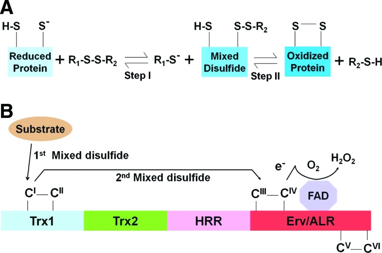
Significance: The cysteine (Cys) residues of proteins play two fundamentally important roles. They serve as sites of post-translational redox modifications as well as influence the conformation of the protein through the formation of disulfide bonds.

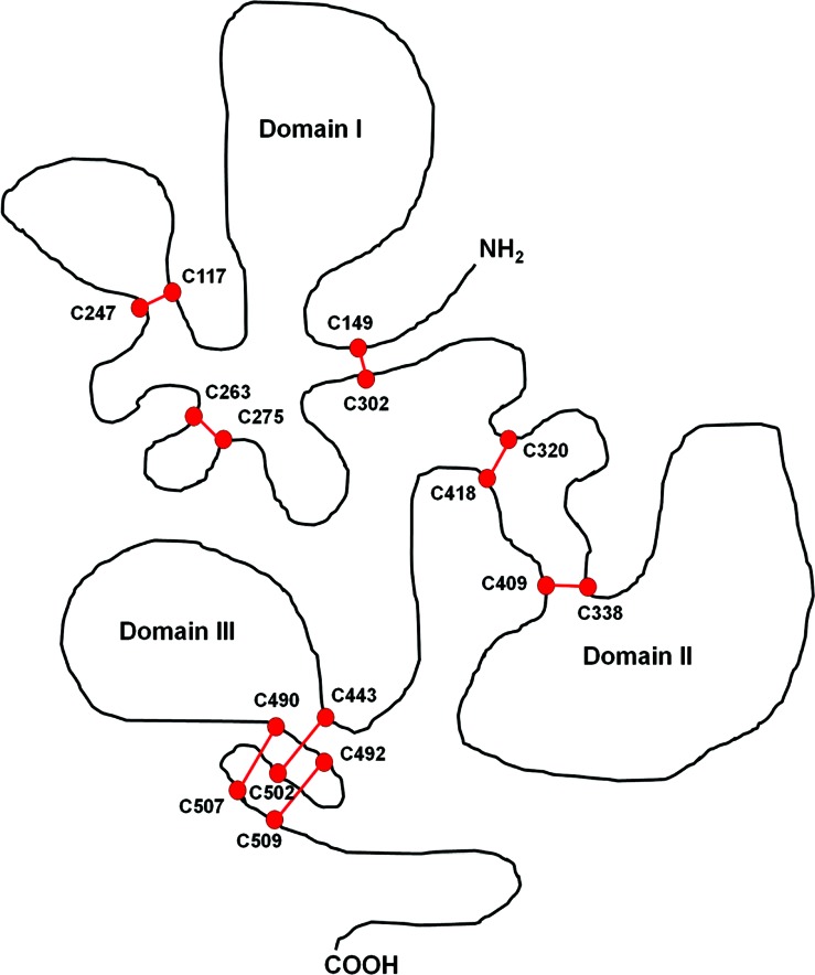
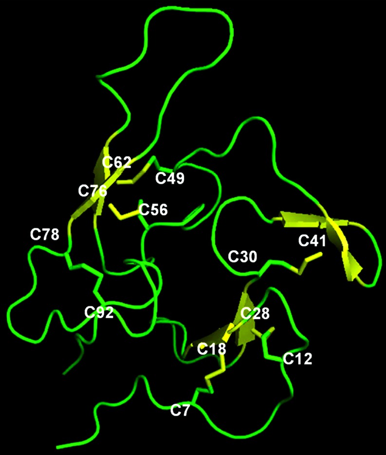
Recent advances: Redox-related and redox-associated protein folding in protozoan parasites has been found to be a major mode of regulation, affecting myriad aspects of the parasitic life cycle, host-parasite interactions, and the disease pathology. Available genome sequences of various parasites have begun to complement the classical biochemical and enzymological studies of these processes. In this article, we summarize the reversible Cys disulfide (S-S) bond formation in various classes of strategically important parasitic proteins, and its structural consequence and functional relevance.

Critical issues: Molecular mechanisms of folding remain under-studied and often disconnected from functional relevance.

Future directions: The clinical benefit of redox research will require a comprehensive characterization of the various isoforms and paralogs of the redox enzymes and their concerted effect on the structure and function of the specific parasitic client proteins.


Figures

FIG. 1.
FIG. 1.
Schematic of redox reactions. (A) Representative thiol-disulfide exchange reaction between a protein and an electron acceptor reagent. This two-step reaction between the redox reagent (R1-S-S-R2) and a substrate protein leads to a disulfide bond formation in the protein and reduction of the electron acceptor. (B) Flow of reducing equivalents through the structural domains of a metazoan Quiescin-sulfhydryl oxidase during catalysis. The thiroedoxin (Trx), Helix-rich region (HRR) and the Erv/ALR domains, and the six Cys residues (CI–CVI) have been described in the text. (To see this illustration in color, the reader is referred to the web version of this article at www.liebertonline.com/ars). Cys, cysteine.
FIG. 2.
FIG. 2.
Schematic of the major domains of plasmodial PDI enzymes. Pf=Plasmodium falciparum, Pb=Plasmodium berghei, Pk=Plasmodium knowlesi, Pv=Plasmodium vivax, Py=Plasmodium yoelii. The suffix numbers (e.g., PfPDI-8) refer to the number of the known chromosomes in which the protein disulfide isomerase gene is located. The GenBank accession numbers are, from the top, NP_704277, NP_704733, NP_701212, NP_702583, DQ266891, DQ266890, DQ266892, and EAA17481. The number of amino-acid residues in each polypeptide is shown on the right. The boxed domains with their catalytic CXXC motif are color-coded. The a-b-b'-a' arrangement of the domains are also highlighted in color, whereby a and a' are the N- and C-terminal catalytic domains, and b, b' are catalytically inactive. We refrained from applying the letter designation to PfPDI-11 due to the relatively central position of its single CGHS domain and the lack of knowledge of its catalytic activity. The N-terminal signal peptide (Blue) and C-terminal ER retention sequences (tetrapeptides) are indicated. The drawing scale is approximate. (To see this illustration in color, the reader is referred to the web version of this article at www.liebertonline.com/ars). CXXC, Cys-X-X-Cys; CGHS, Cys-Gly-His-Cys; ER, endoplasmic reticulum; PDI, protein disulfide isomerase.
FIG. 3.
FIG. 3.
A schematic representation of eight Cys pairs of PfAMA-1 bridged by disulfide bonds. The Cys residues (C with numbers) and the S-S bridges are indicated in red. The three domains are shown as I, II, and II (9, 44, 64, 70). Note that the disulfide bridges generate proper folding of the protein. (To see this illustration in color, the reader is referred to the web version of this article at www.liebertonline.com/ars).
FIG. 4.
FIG. 4.
Schematic diagram of the PfEMP1 polypeptide. The relevant domains are shown in grayscale codes and detailed in the text. The seven key Cys residues of the DBL1α domain involved in disulfide bridge formation are indicated in the expanded view. (To see this illustration in color, the reader is referred to the web version of this article at www.liebertonline.com/ars).
FIG. 5.
FIG. 5.
Crystal structure of PfMSP-1, showing the S-S bridged Cys pairs. The structure is based on the solution NMR structure of PfMSP-1 (NCBI 1CEJ) (47). The beta-strands are yellow, and the loops are green. The six Cys (C) disulfide pairs are C7-C18, C12-C28, C30-C41, C49-C62, C56-C76, and C78-C92. The pairs are shown in a stick model colored by their location in the secondary structure. (To see this illustration in color, the reader is referred to the web version of this article at www.liebertonline.com/ars). This and all other structures in this article were drawn by PyMOL (24).
FIG. 6.
FIG. 6.
Cys disulfide formation causes rotation of amino-acid side chains. In Schistosoma cyclophiln A (CyPA), the two closely spaced Cys residues, Cys122 and Cys126, undergo reversible oxidation reduction. This does not appreciably change the secondary structural elements of the protein, as shown by the perfect superimposition of the two structures, with NCBI PDB ID 2CK1 (oxidized) and 2CMT (reduced) (Panel A). However, a number of amino-acid side chains shifted positions, which includes the active site residue Arg62 (yellow in the oxidized, pink in the reduced structure). A few other rotated side chains are also illustrated in contrasting colors (Panel B). (To see this illustration in color, the reader is referred to the web version of this article at www.liebertonline.com/ars).
FIG. 7.
FIG. 7.
Two representative Cys pairs in the corresponding positions of two Tb VSG primary structures. The VSG names are shown on the left. Horizontal brackets indicate the two S-S bridges in blue (Cys15-Cys145) and red (Cys123-Cys187), respectively (6, 14). The corresponding crystal structures are shown in Figure 8. (To see this illustration in color, the reader is referred to the web version of this article at www.liebertonline.com/ars). VSG, variable surface glycoprotein.
FIG. 8.
FIG. 8.
X-ray diffraction crystal structures of two representative Tb VSG. Ribbon diagram of the crystal structures shows the similar locations of a set of bridged Cys pairs in the two higher order structures even though the primary structures were substantially dissimilar due to the need for antigenic variability in the VSG proteins (12, 17, 35). These and other Cys-Cys disulfide bridges (pair of oxidized Cys joined by disulfide bridge [S-S]) play a crucial role in imparting the native conformation to VSG. The PDB numbers of the two structures (at NCBI) are 1VSG (MITat1.2) and 2VSG (ILTat 1.24). A line drawing of the same two polypeptides is shown in Figure 7. (To see this illustration in color, the reader is referred to the web version of this article at www.liebertonline.com/ars).

References

    1. Adams B. Musiyenko A. Kumar R. Barik S. A novel class of dual-family immunophilins. J Biol Chem. 2005;280:24308–24314. - PMC - PubMed
    1. Adisa A. Rug M. Foley M. Tilley L. Characterisation of a delta-COP homologue in the malaria parasite, Plasmodium falciparum. Mol Biochem Parasitol. 2002;123:11–21. - PubMed
    1. Agorio A. Chalar C. Cardozo S. Salinas G. Alternative mRNAs arising from trans- splicing code for mitochondrial and cytosolic variants of Echinococcus granulosus thioredoxin glutathione reductase. J Biol Chem. 2003;278:12920–12928. - PubMed
    1. Aguilar-Díaz H. Carrero JC. Argüello-García R. Laclette JP. Morales-Montor J. Cyst and encystment in protozoan parasites: optimal targets for new life-cycle interrupting strategies? Trends Parasitol. 2011;27:450–458. - PubMed
    1. Alger HM. Williams DL. The disulfide redox system of Schistosoma mansoni and the importance of a multifunctional enzyme, thioredoxin glutathione reductase. Mol Biochem Parasitol. 2002;121:129–139. - PubMed

Publication types

LinkOut - more resources