

Cone beam computed tomography and intraoral radiography for diagnosis of dental abnormalities in dogs and cats
- PMID: 22122905
- PMCID: PMC3232399
- DOI: 10.4142/jvs.2011.12.4.387
Cone beam computed tomography and intraoral radiography for diagnosis of dental abnormalities in dogs and cats
Abstract
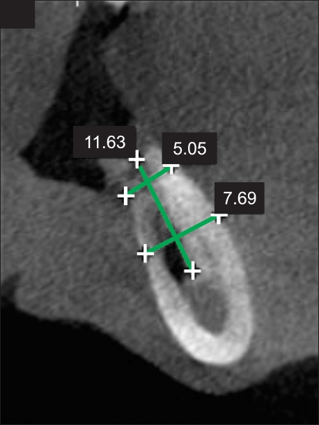
The development of veterinary dentistry has substantially improved the ability to diagnose canine and feline dental abnormalities. Consequently, examinations previously performed only on humans are now available for small animals, thus improving the diagnostic quality. This has increased the need for technical qualification of veterinary professionals and increased technological investments. This study evaluated the use of cone beam computed tomography and intraoral radiography as complementary exams for diagnosing dental abnormalities in dogs and cats. Cone beam computed tomography was provided faster image acquisition with high image quality, was associated with low ionizing radiation levels, enabled image editing, and reduced the exam duration. Our results showed that radiography was an effective method for dental radiographic examination with low cost and fast execution times, and can be performed during surgical procedures.
Figures


Similar articles
-
Diagnostic Imaging of Oral and Maxillofacial Anatomy and Pathology.Vet Clin North Am Small Anim Pract. 2022 Jan;52(1):67-105. doi: 10.1016/j.cvsm.2021.08.003. Vet Clin North Am Small Anim Pract. 2022. PMID: 34838256 Review.
-
Clinical feline dental radiography.Vet Clin North Am Small Anim Pract. 2013 May;43(3):533-554. doi: 10.1016/j.cvsm.2013.02.003. Epub 2013 Mar 30. Vet Clin North Am Small Anim Pract. 2013. PMID: 23643020 Review.
-
Clinical canine dental radiography.Vet Clin North Am Small Anim Pract. 2013 May;43(3):507-532. doi: 10.1016/j.cvsm.2013.02.011. Vet Clin North Am Small Anim Pract. 2013. PMID: 23643019 Review.
-
Dental radiographic interpretation.J Vet Dent. 2005 Mar;22(1):53-9. J Vet Dent. 2005. PMID: 15909456 Review. No abstract available.
-
Problems associated with veterinary dental radiography.Probl Vet Med. 1990 Mar;2(1):46-84. Probl Vet Med. 1990. PMID: 2134590 Review.
Cited by
-
Cone-beam computed tomography of the head in standing equids.BMC Vet Res. 2019 Aug 13;15(1):289. doi: 10.1186/s12917-019-2045-z. BMC Vet Res. 2019. PMID: 31409395 Free PMC article.
-
Prevalence of ear disease in cats undergoing cone beam computed tomography for dental procedures.Front Vet Sci. 2025 Mar 14;12:1553585. doi: 10.3389/fvets.2025.1553585. eCollection 2025. Front Vet Sci. 2025. PMID: 40191083 Free PMC article.
-
The use of micro-computed tomography in the diagnosis of dental and oral disease in rabbits.BMC Vet Res. 2014 Sep 5;10:209. doi: 10.1186/s12917-014-0209-4. BMC Vet Res. 2014. PMID: 25189123 Free PMC article.
-
Diagnostic Yield of Dental Radiography and Cone-Beam Computed Tomography for the Identification of Anatomic Structures in Cats.Front Vet Sci. 2019 Feb 28;6:58. doi: 10.3389/fvets.2019.00058. eCollection 2019. Front Vet Sci. 2019. PMID: 30873423 Free PMC article.
-
Effectiveness of a Nanohydroxyapatite-Based Hydrogel on Alveolar Bone Regeneration in Post-Extraction Sockets of Dogs with Naturally Occurring Periodontitis.Vet Sci. 2021 Dec 26;9(1):7. doi: 10.3390/vetsci9010007. Vet Sci. 2021. PMID: 35051091 Free PMC article.
References
-
- Cohenca N, Simon JH, Roges R, Morag Y, Malfaz JM. Clinical indications for digital imaging in dento-alveolar trauma. Part 1: traumatic injuries. Dent Traumatol. 2007;23:95–104. - PubMed
-
- Costa MAF, Costa MFB, Roza MR, Gama Filho JB. Biossegurança em odontologia veterinária. In: Roza MR, editor. Odontologia em Pequenos Animais. Rio de Janeiro: LF Livros de Veterinária; 2004. pp. 19–38.
-
- Farman AG, Scarfe WC. Development of imaging selection criteria and procedures should precede cephalometric assessment with cone-beam computed tomography. Am J Orthod Dentofacial Orthop. 2006;130:257–265. - PubMed
-
- Fullmer JM, Scarfe WC, Kushner GM, Albert B, Farman AG. Cone beam computed tomographic findings in refractory chronic suppurative osteomyelitis of the mandible. Br J Oral Maxillofac Surg. 2007;45:364–371. - PubMed
-
- Garib DG, Raymundo R, Jr, Raymundo MV, Raymundo DV, Ferreira SN. Tomografia computadorizada de feixe cônico (Cone Beam): Entendendo este novo método de diagnóstico por imagem com promissora aplicabilidade na ortodontia. Rev Dent Press Ortodon Ortop Facial. 2007;12:139–156.
Publication types
MeSH terms
Substances
LinkOut - more resources
Full Text Sources
Medical
Miscellaneous
